J Vet Sci.
2011 Dec;12(4):387-392. 10.4142/jvs.2011.12.4.387.
Cone beam computed tomography and intraoral radiography for diagnosis of dental abnormalities in dogs and cats
- Affiliations
-
- 1Department of Veterinary Medicine, Federal University of Goias, Goiania 74001-970, Brazil. marcelloroza@gmail.com
- 2Department of Radiology, Faculty of Dentistry, Catholic University of Brasilia, Brasilia 71966-700, Brazil.
- 3International Team for Implantology, Sapound;o Paulo 04551-060, Brazil.
- 4Faculty of Health Sciences, University of Brasilia, Brasilia 70910-700, Brazil.
- KMID: 1365023
- DOI: http://doi.org/10.4142/jvs.2011.12.4.387
Abstract
- The development of veterinary dentistry has substantially improved the ability to diagnose canine and feline dental abnormalities. Consequently, examinations previously performed only on humans are now available for small animals, thus improving the diagnostic quality. This has increased the need for technical qualification of veterinary professionals and increased technological investments. This study evaluated the use of cone beam computed tomography and intraoral radiography as complementary exams for diagnosing dental abnormalities in dogs and cats. Cone beam computed tomography was provided faster image acquisition with high image quality, was associated with low ionizing radiation levels, enabled image editing, and reduced the exam duration. Our results showed that radiography was an effective method for dental radiographic examination with low cost and fast execution times, and can be performed during surgical procedures.
MeSH Terms
Figure
-

Fig. 1 Cat placed under cone beam computed tomography (CBCT) scanner via a polyvinyl chloride device.
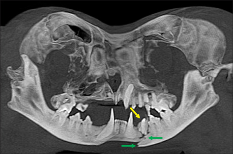
Fig. 2 Cone beam CT of a cat's head after trauma. Panoramic view (bidimensional recontruction) showing mandibular (green arrows) and dental (yellow arrow) fractures.
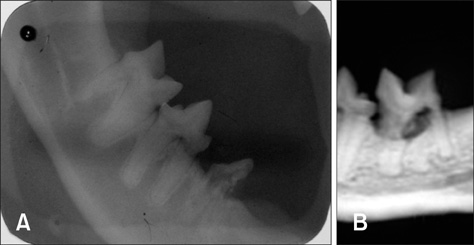
Fig. 3 Image of a feline dental resorption lesion. (A) Intraoral radiography (IOR) and (B) CBCT.
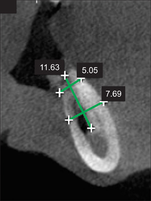
Fig. 4 Oblique view of a transverse CBCT section showing the mandibular area suitable for dental implantation. The software allowed measurement of the bone that helps determine the choice of implant.

Fig. 5 IOR showing dental implant in the mandibular area.
Reference
-
1. Cohenca N, Simon JH, Roges R, Morag Y, Malfaz JM. Clinical indications for digital imaging in dento-alveolar trauma. Part 1: traumatic injuries. Dent Traumatol. 2007. 23:95–104.
Article2. Costa MAF, Costa MFB, Roza MR, Gama Filho JB. Roza MR, editor. Biossegurança em odontologia veterinária. Odontologia em Pequenos Animais. 2004. Rio de Janeiro: LF Livros de Veterinária;19–38.3. Farman AG, Scarfe WC. Development of imaging selection criteria and procedures should precede cephalometric assessment with cone-beam computed tomography. Am J Orthod Dentofacial Orthop. 2006. 130:257–265.
Article4. Fullmer JM, Scarfe WC, Kushner GM, Albert B, Farman AG. Cone beam computed tomographic findings in refractory chronic suppurative osteomyelitis of the mandible. Br J Oral Maxillofac Surg. 2007. 45:364–371.
Article5. Garib DG, Raymundo R Jr, Raymundo MV, Raymundo DV, Ferreira SN. Tomografia computadorizada de feixe cônico (Cone Beam): Entendendo este novo método de diagnóstico por imagem com promissora aplicabilidade na ortodontia. Rev Dent Press Ortodon Ortop Facial. 2007. 12:139–156.
Article6. Guerrero ME, Jacobs R, Loubele M, Schutyser F, Suetens P, van Steenberghe D. State-of-the-art on cone beam CT imaging for preoperative planning of implant placement. Clin Oral Investig. 2006. 10:1–7.
Article7. Hatcher DC, Aboudara CL. Diagnosis goes digital. Am J Orthod Dentofacial Orthop. 2004. 125:512–515.
Article8. Honey OB, Scarfe WC, Hilgers MJ, Klueber K, Silveira AM, Haskell BS, Farman AG. Accuracy of cone-beam computed tomography imaging of the temporomandibular joint: comparisons with panoramic radiology and linear tomography. Am J Orthod Dentofacial Orthop. 2007. 132:429–438.
Article9. Iplikçioğlu H, Akça K, Çehreli MC. The use of computerized tomography for diagnosis and treatment planning in implant dentistry. J Oral Implantol. 2002. 28:29–36.
Article10. Kau CH, Richmond S, Palomo JM, Hans MG. Three-dimensional cone beam computerized tomography in orthodontics. J Orthod. 2005. 32:282–293.11. Mah JK, Danforth RA, Bumann A, Hatcher D. Radiation absorbed in maxillofacial imaging with a new dental computed tomography device. Oral Surg Oral Med Oral Pathol Oral Radiol Endod. 2003. 96:508–513.
Article12. Mozzo P, Procacci C, Tacconi A, Martini PT, Andreis IAB. A new volumetric CT machine for dental imaging based on the cone-beam technique: preliminary results. Eur Radiol. 1998. 8:1558–1564.
Article13. Natalini CC. Teoria e Técnicas em Anestesiologia Veterinária. 2007. Porto Alegre: Artmed;83–88.14. Negro VB, Hernández SZ, Saccomanno DM. Detección de lesiones odontoclásticas reabsortivas felinas (LORF) mediante examen clínico y radiológico. In Vet. 2005. 7:87–97.15. Niemiec BA, Furman R. Canine dental radiography. J Vet Dent. 2004. 21:186–190.16. Niemiec BA, Furman R. Feline dental radiography. J Vet Dent. 2004. 21:252–257.17. Roza MR. Odontologia em Pequenos Animais. 2004. Rio de Janeiro: LF Livros de Veterinária;119–136.18. Roza MR, Gama Filho JB, Costa MAF. Biossegurança em Ambientes Hospitalares Veterinários. 2003. Rio de Janeiro: Interciência;43–58.19. Roza MR, Silva LAF, Januário AL, Barriviera M, Oliveira ACA, Fioravanti MCS. Tomografia computadorizada de feixe cônico na odontologia veterinária: descrição e padronização da técnica. Pesqui Vet Bras. 2009. 29:617–624.
Article20. Scarfe WC, Farman AG, Sukovic P. Clinical applications of cone-beam computed tomography in dental practice. J Can Dent Assoc. 2006. 72:75–80.21. Scarfe WC. Imaging of maxillofacial trauma: evolutions and emerging revolutions. Oral Surg Oral Med Oral Pathol Oral Radiol Endod. 2005. 100:2 Suppl. S75–S96.
Article22. Tsugawa AJ, Verstraete FJM. How to Obtain and Interpret Periodontal Radiographs in Dogs. Clin Tech Small Anim Pract. 2000. 15:204–210.
Article23. Turkyilmaz I, Tözüm TF, Tumer C. Bone density assessments of oral implant sites using computerized tomography. J Oral Rehabil. 2007. 34:267–272.
Article24. van Wessum R, Harvey CE, Hennet P. Feline dental resorptive lesions. Prevalence patterns. Vet Clin North Am Small Anim Pract. 1992. 22:1405–1416.
Article25. Verstraete FJ, Kass PH, Terpak CH. Diagnostic value of full-mouth radiography in cats. Am J Vet Res. 1998. 59:692–695.26. Winter AA, Pollack AS, Frommer HH, Koenig L. Cone Beam volumetric tomography vs. medical CT scanners: Expanding dental applications. NY State Dent J. 2005. 71:28–33.27. Yoshikawa H, Watanabe K, Ozawa T. Odontoclastic resorptive lesions in a dog. J Vet Med Sci. 2008. 70:103–105.
Article
- Full Text Links
-
- Actions
-
Cited
- CITED
-
- Close
- Share
-
- Similar articles
-
- Detection of maxillary second molar with two palatal roots using cone beam computed tomography: a case report
- Three-dimensional imaging modalities in endodontics
- Basic principle of cone beam computed tomography
- A rare case of dilated invaginated odontome with talon cusp in a permanent maxillary central incisor diagnosed by cone beam computed tomography
- New evolution of cone-beam computed tomography in dentistry: Combining digital technologies
