Spontaneous Regression of Sclerosing Mesenteritis Presenting as a Huge Mass
- Affiliations
-
- 1Department of Internal Medicine, The Catholic University of Korea, College of Medicine, Seoul, Korea. jikim@catholic.ac.kr
- 2Department of Hostipal Pathology, The Catholic University of Korea, College of Medicine, Seoul, Korea.
- KMID: 1364837
- DOI: http://doi.org/10.4166/kjg.2012.59.4.317
Abstract
- Sclerosing mesenteritis is a rare benign disease originated from the mesenteries. It can be related to autoimmune disease, vasculitis, ischemia, infection, trauma and operation, but most of cases are idiopathic. The overall prognosis of sclerosing mesenteritis is usually good with benign, course. However, no consensus of treatment has yet been established. We report a case of spontaneous partial regression of sclerosing mesenteritis presented as a huge mass and diagnosed by finding of contrast enhanced abdominal computed tomography and percutaneous ultrasonography guided needle biopsy.
MeSH Terms
Figure
-
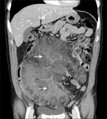
Fig. 1 Abdomen computed tomography image with intravenous contrast showed huge mass like lesion with congestion of the mesenteric vessel (arrows).

Fig. 2 Small bowel series showed the displacement of the small bowel due to mesenteric mass.

Fig. 3 Microscopic finding showed focal infiltration of chronic inflammatory cells and fibroblast (H&E, ×100).

Fig. 4 Follow up abdomen computed tomography performed 3 months later showed much regression of previously noted mass like lesion of the mesenteries.
Cited by 2 articles
-
A Case of IgG4-Related Sclerosing Mesenteritis Associated with Crohn's Disease
Eui Jung Kim, Eun Young Kim, Jung Eun Song, Hyeon Chul Lee, Gyu Hwan Bae, Hoon Kyu Oh, Tae Sung Lee
Korean J Gastroenterol. 2014;63(3):176-182. doi: 10.4166/kjg.2014.63.3.176.Immunoglobulin G4 Unrelated Idiopathic Mesenteric Sclerosis
Tae Hyung Kwon, Kwang Bum Cho, Hyun Jik Lee, Sun Young Kwon, Yoon Suk Lee
Korean J Gastroenterol. 2019;73(1):50-55. doi: 10.4166/kjg.2019.73.1.50.
Reference
-
1. Emory TS, Monihan JM, Carr NJ, Sobin LH. Sclerosing mesenteritis, mesenteric panniculitis and mesenteric lipodystrophy: a single entity? Am J Surg Pathol. 1997. 21:392–398.2. Cuff R, Landercasper J, Schlack S. Sclerosing mesenteritis. Surgery. 2001. 129:509–510.3. Bush RW, Hammar SP Jr, Rudolph RH. Sclerosing mesenteritis. Response to cyclophosphamide. Arch Intern Med. 1986. 146:503–505.4. Bala A, Coderre SP, Johnson DR, Nayak V. Treatment of sclerosing mesenteritis with corticosteroids and azathioprine. Can J Gastroenterol. 2001. 15:533–535.5. Lee SY, Park DE, Chae KM. Sclerosing mesenteritis. J Korean Surg Soc. 2006. 71:218–221.6. Chawla S, Yalamarthi S, Shaikh IA, Tagore V, Skaife P. An unusual presentation of sclerosing mesenteritis as pneumoperitoneum: Case report with a review of the literature. World J Gastroenterol. 2009. 15:117–120.7. Sabate JM, Torrubia S, Maideu J, Franquet T, Monill JM, Perez C. Sclerosing mesenteritis: imaging findings in 17 patients. AJR Am J Roentgenol. 1999. 172:625–629.8. Delgado Plasencia L, Rodríguez Ballester L, López-Tomassetti Fernández EM, et al. Mesenteric panniculitis: experience in our center. Rev Esp Enferm Dig. 2007. 99:291–297.9. Schaffler A, Herfarth H. Creeping fat in Crohn's disease: travelling in a creeper lane of research? Gut. 2005. 54:742–744.10. Soergel KH, Hensley GT. Fatal mesenteric panniculitis. Gastroenterology. 1966. 51:529–536.11. Jeon EJ, Cho SM. Idiopathic isolated omental panniculitis confirmed by percutaneous CT-guided biopsy. Gut Liver. 2009. 3:321–324.12. Remmele W, Muller-Lobeck H, Paulus W. Primary mesenteritis, mesenteric fibrosis and mesenteric fibromatosis: report of four cases, pathology, and classification. Pathol Res Pract. 1988. 184:77–85.13. Li LY, Cho YK, Lee JH, et al. Two cases of mesenteric panniculitis treated by medical management. Korean J Med. 2006. 71:S934–S939.14. Park KH, Chang HK, Choi SY, et al. Sclerosing mesenteritis associated with skin panniculitis and pleural thickening. Korean J Med. 1999. 57:103–107.15. Min BH, Shinn YC. Idiopathic mesenteric fibrosis. J Korean Surg Soc. 1976. 18:73–76.16. Cha SJ, Linn HM, Chang ST, Part YW, Yoo JH, Song KY. Primary mesenteritis-A case report. J Korean Surg Soc. 1991. 41:819–829.17. Issa I, Baydoun H. Mesenteric panniculitis: Various presentations and treatment regimens. World J Gastroenterol. 2009. 15:3827–3830.18. Daskalogiannaki M, Voloudaki A, Prassopoulos P, et al. CT evaluation of mesenteric panniculitis: prevalence and associated diseases. AJR Am J Roentgenol. 2000. 174:427–431.
- Full Text Links
-
- Actions
-
Cited
- CITED
-
- Close
- Share
-
- Similar articles
-
- Sclerosing Mesenteritis
- Immunoglobulin G4 Unrelated Idiopathic Mesenteric Sclerosis
- A Case of Idiopathic Sclerosing Mesenteritis with Retroperitoneal Fibrosis
- A Case of Sclerosing Mediastinitis Presenting as Esophageal Stricture
- Cholangiocarcinoma with Regional Lymph Node Metastasis Masquerading as Sclerosing Mesenteritis
