Intracranial Extramedullary Hematopoiesis in Beta-Thalassemia
- Affiliations
-
- 1Department of Medical Imaging, Nan fang Hospital, Southern Medical University, Guangzhou, Guangdong 510515, China. daoshi08@gmail.com
- 2Department of Neurosurgery, Zhujiang Hospital, Southern Medical University, Guangzhou, Guangdong 510282, China.
- KMID: 1245390
- DOI: http://doi.org/10.3348/kjr.2012.13.2.240
Abstract
- Extramedullary hematopoiesis (EMH) represents tumor-like proliferation of hemopoietic tissue which complicates chronic hemoglobinopathy. Intracranial EMH is an extremely rare occurrence. Magnetic resonance imaging (MRI) offers a precise diagnosis. It is essential to distinguish EMH from other extradural central nervous system tumors, because treatment and prognosis are totally different. Herein, we report the imaging findings of beta-thalassemia in a 13-year-old boy complaining of weakness of left side of the body and gait disturbance; CT and MRI revealed an extradural mass in the right temporoparietal region.
Keyword
MeSH Terms
Figure
-
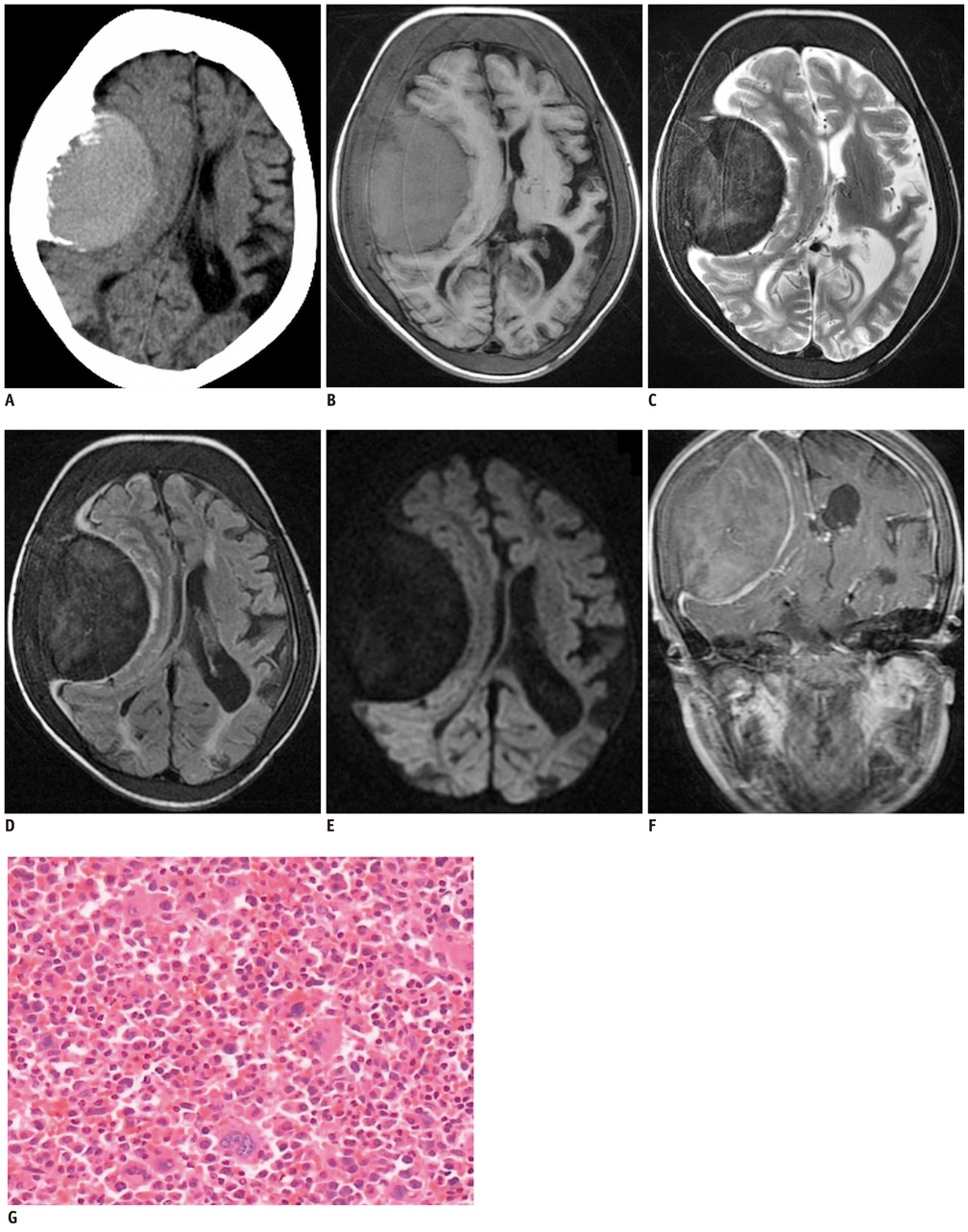
Fig. 1 Intracranial EMH in 13-year-old boy with beta-thalassemia. A. Axial non-contrast CT showing soft tissue mass with density of 45 Hounsfield units in right temporo-parietal region with smooth margin compressing adjacent brain parenchyma. Mass is in close contact with diploic space of skull and displays periosteal reaction. B. T1-weighted MR image showing homogeneously slightly high signal intensity mass compared to gray matter with clear margin in right temporo-parietal region, causing buckling of white matter, ipsilateral ventricle compression, and midline shift. Uniform thickening of diploic space and skull bone is also seen. C-E. Mass showing homogeneously low signal intensity on T2-weighted image (C), fluid attenuation inversion recovery (D) and diffusion weighted imaging (E). F. Contrast enhanced coronal T1-weighted image showing significant dural enhancement and moderate enhancement of mass. G. Pathological examination of postoperative specimen on Hematoxylin and Eosin staining showing round to oval cells with erythroblasts, megakaryocytes and promyelocytes (magnification, × 400).
Reference
-
1. Tsitouridis J, Stamos S, Hassapopoulou E, Tsitouridis K, Nikolopoulos P. Extramedullary paraspinal hematopoiesis in thalassemia: CT and MRI evaluation. Eur J Radiol. 1999. 30:33–38.2. Haidar S, Ortiz-Neira C, Shroff M, Gilday D, Blaser S. Intracranial involvement in extramedullary hematopoiesis: case report and review of the literature. Pediatr Radiol. 2005. 35:630–634.3. Mathews MS, Duma CM, Brant-Zawadzki M, Hasso A, Westhout FD, Klein DJ, et al. Extramedullary hematopoeisis within a convexity meningioma. Surg Neurol. 2008. 69:522–525. discussion 525.4. Koch BL, Bisset GS 3rd, Bisset RR, Zimmer MB. Intracranial extramedullary hematopoiesis: MR findings with pathologic correlation. AJR Am J Roentgenol. 1994. 162:1419–1420.5. Beckner ME, Lee JY, Schochet SS Jr, Chu CT. Intracranial extramedullary hematopoiesis associated with pilocytic astrocytoma: a case report. Acta Neuropathol. 2003. 106:584–587.6. Kang BK, Na DG, Ryoo JW, Byun HS, Roh HG, Pyeun YS. Diffusion-weighted MR imaging of intracerebral hemorrhage. Korean J Radiol. 2001. 2:183–191.7. Klisch J, Husstedt H, Hennings S, von Velthoven V, Pagenstecher A, Schumacher M. Supratentorial primitive neuroectodermal tumours: diffusion-weighted MRI. Neuroradiology. 2000. 42:393–398.8. Smidt MH, de Bruin HG, van't Veer MB, van den Bent MJ. Intracranial granulocytic sarcoma (chloroma) may mimic a subdural hematoma. J Neurol. 2005. 252:498–499.9. Kremer S, Grand S, Rémy C, Pasquier B, Benabid AL, Bracard S, et al. Contribution of dynamic contrast MR imaging to the differentiation between dural metastasis and meningioma. Neuroradiology. 2004. 46:642–648.10. Tun K, Kaptanoglu E, Celikmez RC, Gurcan O, Turkoglu OF, Kutluay L. Meningeal extramedullary haematopoiesis mimicking subdural hematoma. J Clin Neurosci. 2008. 15:208–210.
- Full Text Links
-
- Actions
-
Cited
- CITED
-
- Close
- Share
-
- Similar articles
-
- Extramedullary Hematopoiesis Visualized on FDG‑PET/CT in a Patient with Beta‑Thalassemia
- Periportal Extramedullary Hematopoiesis
- A Case of Extramedullary Hematopoiesis Associated with Congenital Dyserythropoietic Anemia
- Cutaneous Extramedullary Hematopoiesis in Idiopathic Myelofibrosis
- VATS Resection for a Posterior Mediastinal Extramedullary Hematopoietic Mass: Resection of Extramedullary Hematopoiesis
