Korean J Radiol.
2011 Jun;12(3):382-385. 10.3348/kjr.2011.12.3.382.
Primary Hepatic Amyloidosis: Report of an Unusual Case Presenting as a Mass
- Affiliations
-
- 1Department of Radiology, College of Medicine, Yeungnam University, Daegu 705-717, Korea. src2179@hanmail.net
- 2Department of Pathology, College of Medicine, Yeungnam University, Daegu 705-717, Korea.
- KMID: 1122336
- DOI: http://doi.org/10.3348/kjr.2011.12.3.382
Abstract
- Hepatic involvement of amyloidosis is common. Diffuse infiltration with hepatomegaly is a usual radiologic finding of hepatic amyloidosis. To our knowledge, this is the first case of amyloidosis involving the liver that presented as a mass.
Keyword
MeSH Terms
Figure
-
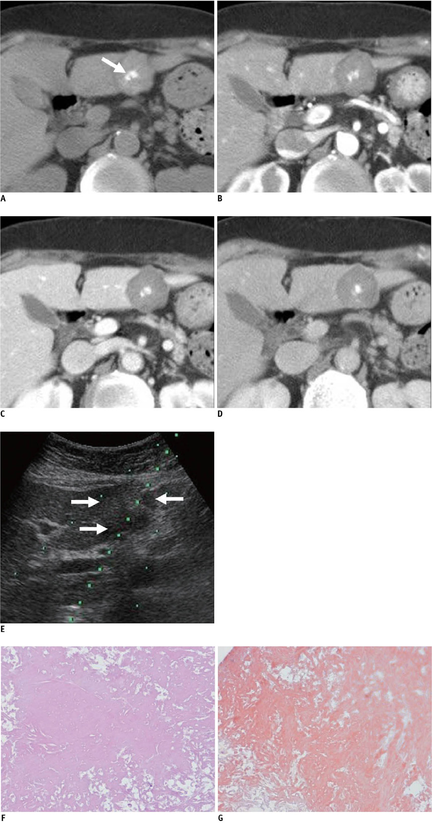
Fig. 1 73-year-old woman with hepatic amyloidosis. A. Unenhanced CT shows high density lobulating mass with central calcification (arrow) in segment 3 of liver. B-D. Mass shows very poor delayed contrast enhancement on arterial (B), portal venous (C) and delayed (D) phase image. E. US image during needle biopsy indicates mild heterogeneous echoic mass on segment 3 of liver (arrows). F, G. Microscopically, diffuse amyloid deposits are present without viable hepatocyte (F, Hematoxylin & Eosin staining, original magnification × 200), and Congo red stain is positive (G).
Reference
-
1. Kim MS, Ryu JA, Park CS, Lee EJ, Park NH, Oh HE, et al. Amyloidosis of the mesentery and small intestine presenting as a mesenteric haematoma. Br J Radiol. 2008. 81:E1–E3.2. Georgiades CS, Neyman EG, Barish MA, Fishman EK. Amyloidosis: review and CT manifestations. Radiographics. 2004. 24:405–416.3. Scott PP, Scott WW Jr, Siegelman SS. Amyloidosis: an overview. Semin Roentgenol. 1986. 21:103–112.4. Glenner GG. Amyloid deposits and amyloidosis. The betafibrilloses (first of two parts). N Engl J Med. 1980. 302:1283–1292.5. Kim SH, Han JK, Lee KH, Won HJ, Kim KW, Kim JS, et al. Abdominal amyloidosis: spectrum of radiological findings. Clin Radiol. 2003. 58:610–620.6. WHO-IUIS Nomenclature Sub-Committee. Nomenclature of amyloid and amyloidosis. Bull World Health Organ. 1993. 71:105–112.7. Gertz MA, Kyle RA. Amyloidosis with IgM monoclonal gammopathies. Semin Oncol. 2003. 30:325–328.8. Joss N, McLaughlin K, Simpson K, Boulton-Jones JM. Presentation, survival and prognostic markers in AA amyloidosis. Qjm. 2000. 93:535–542.9. Pasqualetti P, Casale R. Risk of malignant transformation in patients with monoclonal gammopathy of undetermined significance. Biomed Pharmacother. 1997. 51:74–78.10. Maniatis A. Pathophysiology of paraprotein production. Ren Fail. 1998. 20:821–828.11. Dhodapkar MV, Merlini G, Solomon A. Biology and therapy of immunoglobulin deposition diseases. Hematol Oncol Clin North Am. 1997. 11:89–110.12. Gertz MA, Kyle RA. Hepatic amyloidosis: clinical appraisal in 77 patients. Hepatology. 1997. 25:118–121.13. Gastineau DA, Gertz MA, Rosen CB, Kyle RA. Computed tomography for diagnosis of hepatic rupture in primary systemic amyloidosis. Am J Hematol. 1991. 37:194–196.14. Monzawa S, Tsukamoto T, Omata K, Hosoda K, Araki T, Sugimura K. A case with primary amyloidosis of the liver and spleen: radiologic findings. Eur J Radiol. 2002. 41:237–241.15. Suzuki S, Takizawa K, Nakajima Y, Katayama M, Sagawa F. CT findings in hepatic and splenic amyloidosis. J Comput Assist Tomogr. 1986. 10:332–334.16. Kennan NM, Evans C. Case report: hepatic and splenic calcification due to amyloid. Clin Radiol. 1991. 44:60–61.17. Jacobs JE, Birnbaum BA, Furth EE. Abdominal visceral calcification in primary amyloidosis: CT findings. Abdom Imaging. 1997. 22:519–521.18. Benson L, Hemmingsson A, Ericsson A, Jung B, Sperber G, Thuomas KA, et al. Magnetic resonance imaging in primary amyloidosis. Acta Radiol. 1987. 28:13–15.19. Coumbaras M, Chopier J, Massiani MA, Antoine M, Boudghene F, Bazot M. Diffuse mesenteric and omental infiltration by amyloidosis with omental calcification mimicking abdominal carcinomatosis. Clin Radiol. 2001. 56:674–676.
