Diffuse Hepatic Hemangiomatosis without Extrahepatic Involvement in an Adult Patient
- Affiliations
-
- 1Department of Radiology, St. Vincent's Hospital, College of Medicine, The Catholic University of Korea, Gyeonggi-do, Korea. sshwang7@catholic.ac.kr
- KMID: 1118879
- DOI: http://doi.org/10.3348/kjr.2008.9.6.559
Abstract
- We report an extremely rare case of a diffuse hepatic hemangiomatosis without extrahepatic involvement in an adult. The imaging findings of this tumor were similar to those of a hepatic hemangioma and included contrast enhancement with a centripetal filling pattern of the entire hepatic tumor on the delayed phase of a dynamic CT and inhomogeneous diffuse uptake of the entire tumor on blood-pool images obtained five hours later on a 99mTc-labeled red blood cell scan. Despite its rarity, diffuse hepatic hemangiomatosis can be suggested in adult patients with diffusely involved hepatic tumors showing the radiological findings of a hepatic hemangioma.
Keyword
MeSH Terms
Figure
-
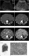
Fig. 1 Diffuse hepatic hemangiomatosis in 33-year-old woman. A. Ultrasonography indicates heterogeneous infiltrative hepatic tumor containing various-sized hypoechoic nodules. B. No remarkable tumor vascularity is observed, except for hepatic parenchymal vessels. C-F. Dynamic CT demonstrates early nodular contrast enhancement on arterial (D) and gradual centripetal pattern of contrast enhancement in diffuse infiltrating hepatic mass on portal (E) and delayed (F) phases. Neither intratumoral calcifications nor area of normalappearing intervening hepatic parenchymal vessels are visible. G. 99mTc-labeled red blood cell scan reveals heterogeneous uptake of radiopharmaceuticals throughout hepatic mass, involving whole liver on blood pool images. Diffuse tumor uptake is still noted on delayed scan obtained five hours later. H. Histological findings reveal endothelial cell proliferation and dilated blood channels (Hematoxylin & Eosin staining, × 200).
Reference
-
1. Lopriore E, Markhorst DG. Diffuse neonatal haemangiomatosis: new views on diagnostic criteria and prognosis. Acta Paediatr. 1999. 88:93–97.2. Lehmann FS, Beglinger C, Schnabel K, Terracciano L. Progressive development of diffuse liver hemangiomatosis. J Hepatol. 1999. 30:951–954.3. Moon WS, Yu HC, Lee JM, Kang MJ. Diffuse hepatic hemangiomatosis in an adult. J Korean Med Sci. 2000. 15:471–474.4. Langner C, Thonhofer R, Hegenbarth K, Trauner M. Diffuse hemangiomatosis of the liver and spleen in an adult. Pathologe. 2001. 22:424–428.5. Latifi HR, Siegel MJ. Diffuse neonatal hemangiomatosis: CT findings in an adult. J Comput Assist Tomogr. 1992. 16:971–973.6. Ohnishi S, Miyagishima T, Nakagawa M, Kamata T, Kishimoto A, Choi GH, et al. Diffuse neonatal hemangiomatosis without cutaneous lesions in an adult-a case report. Angiology. 2002. 53:235–237.7. Bishop PR, Nowicki MJ, Parker PH. Radiological case of the month. Hepatic hemangiomatosis. Arch Pediatr Adolesc Med. 2000. 154:743–774.8. Feurle GE. Arteriovenous shunting and cholestasis in hepatic hemangiomatosis associated with metoclopramide. Gastroenterology. 1990. 99:258–262.9. Buetow PC, Buck JL, Ros PR, Goodman ZD. Malignant vascular tumors of the liver: radiologic-pathologic correlation. Radiographics. 1994. 14:153–166.10. Conter RL, Longmire WP Jr. Recurrent hepatic hemangiomas. Possible association with estrogen therapy. Ann Surg. 1988. 207:115–119.11. Frangides C, Kounis NG, Papadaki PJ, Goudevenos J, Zabras GM. Diffuse hepatic hemangiomatosis in the elderly. Br J Clin Pract. 1995. 49:215–216.
- Full Text Links
-
- Actions
-
Cited
- CITED
-
- Close
- Share
-
- Similar articles
-
- Diffuse hepatic hemangiomatosis in an adult
- Diffuse Neonatal Hemangiomatosis
- Eruptive Neonatal Hemangiomatosis
- Giant cavernous hemangioma coexistent with diffuse hepatic hemangiomatosis presenting as portal vein thrombosis and hepatic lobar atrophy
- Diffuse Neonatal Hemangiomatosis Successfully Treated with High Dose Corticosteroid
