Korean J Radiol.
2004 Dec;5(4):274-279. 10.3348/kjr.2004.5.4.274.
MR Imaging Findings of Painful Type II Accessory Navicular Bone: Correlation with Surgical and Pathologic Studies
- Affiliations
-
- 1Department of Diagnostic Radiology, Eulji Hospital, Eulji University School of Medicine, Korea. cys0128@eulji.or.kr
- 2Department of Orthopedic Surgery, Eulji Hospital, Eulji University School of Medicine, Korea.
- 3Department of Diagnostic Radiology, Seoul National University Bundang Hospital, Korea.
- 4Department of Pathology, Eulji Hospital, Eulji University School of Medicine, Korea.
- KMID: 1118835
- DOI: http://doi.org/10.3348/kjr.2004.5.4.274
Abstract
OBJECTIVE
To evaluate the MR imaging findings of painful type II accessory navicular bone and to correlate these with the surgical and pathologic findings. MATERIALS AND METHODS: The MR images of 17 patients with medial foot pain and surgically proven type II accessory navicular abnormalities were reviewed. The changes of signal intensity in the accessory navicular, synchondrosis and adjacent soft tissue, the presence of synchondrosis widening, and posterior tibial tendon (PTT) pathology on the T1-weighted and fat-suppressed T2-weighted images were analyzed. The MR imaging findings were compared with the surgical and pathologic findings. RESULTS: The fat-suppressed T2-weighted images showed high signal intensity in the accessory navicular bones and synchondroses in all patients, and in the soft tissue in 11 (64.7%) of the 17 patients, as well as synchondrosis widening in 3 (17.6%) of the 17 patients. The MR images showed tendon pathology in 12 (75%) of the 16 patients with PTT dysfunction at surgery. The pathologic findings of 16 surgical specimens included areas of osteonecrosis with granulomatous inflammation, fibrosis and destruction of the cartilage cap. CONCLUSION: The MR imaging findings of painful type II accessory navicular bone are a persistent edema pattern in the accessory navicular bone and within the synchondrosis, indicating osteonecrosis, inflammation and destruction of the cartilage cap. Posterior tibial tendon dysfunction was clinically evident in most patients.
Keyword
MeSH Terms
Figure
-
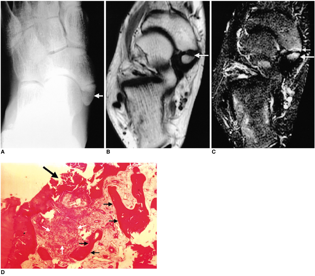
Fig. 1 Painful accessory navicular bone in a 14-year-old male soccer enthusiast.
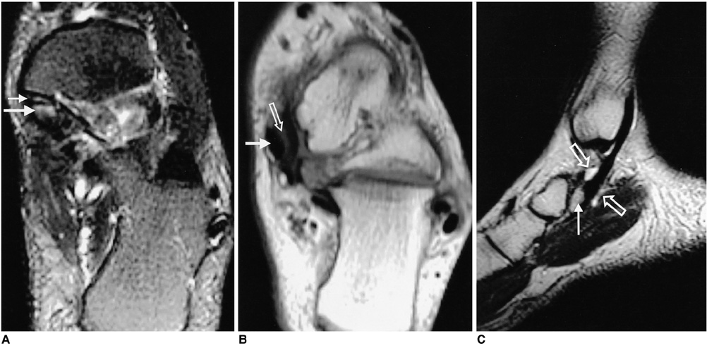
Fig. 2 Surgically proven synovitis with painful accessory navicular bone in a 16-year-old boy.
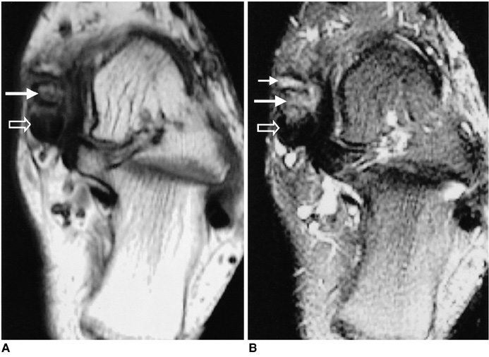
Fig. 3 Surgically proven partial tear of the posterior tibial tendon with painful accessory navicular bone in a 51-year-old woman.
Reference
-
1. Geist ES. Supernumerary bone of the foot: A roentgen study of the feet of 100 normal individuals. Am J Orthop Surg. 1914. 12:403.2. Kruse RW, Chen J. Accessory bones of the foot: clinical significance. Mil Med. 1995. 160:464–467.3. Lawson JP, Ogden JA, Sella E, Barwick KW. The painful accessory navicular. Skeletal Radiol. 1984. 13:250–262.4. Sella EJ, Lawson JP, Ogden JA. The accessory navicular synchondrosis. Clin Orthop. 1986. 209:280–285.5. Miller TT, Staron RB, Feldman F, Parisien M, Glucksman WJ, Gandolfo LH. The symptomatic accessory tarsal navicular bone: assessment with MR imaging. Radiology. 1995. 195:849–853.6. Demeyere N, De Maeseneer M, Osteaux M. Quiz case. Symptomatic type II accessory navicular. Eur J Radiol. 2001. 37:60–63.7. Romanowski CA, Barrington NA. The accessory navicular-an important cause of medial foot pain. Clin Radiol. 1992. 46:261–264.8. Mosel LD, Kat E, Voyvodic F. Imaging of the symptomatic type II accessory navicular bone. Australas Radiol. 2004. 48:267–271.9. Shah S, Achong DM. The painful accessory navicular bone: scintigraphic and radiographic correlation. Clin Nucl Med. 1999. 24:125–126.10. Grogan DP, Gasser SI, Ogden JA. The painful accessory navicular: a clinical and histopathological study. Foot Ankle. 1989. 10:164–169.11. Sella EJ, Lawson JP. Biomechanics of the accessory navicular synchondrosis. Foot Ankle. 1987. 8:156–163.12. Chen YJ, Hsu RW, Liang SC. Degeneration of the accessory navicular synchondrosis presenting as a rupture of the posterior tibial tendon. J Bone Joint Surg Am. 1997. 79:1791–1798.13. Kidner FC. The prehallux in its relation to flat-foot. J Bone Joint Surg Am. 1929. 11:831.14. Chater EH. Foot pain and the accessory navicular bone. Irish J Med Sci. 1962. 442:471–475.15. Schweitzer ME, Caccese R. Karasick D, Wapner KL, Mitchell DG. Posterior tibial tendon tears: utility of secondary signs for MR-imaging diagnosis. Radiology. 1993. 188:655–659.16. Chen YJ, Shih HN, Huang TJ, Hsu RW. Posterior tibial tendon tear combined with a fracture of the accessory navicular: a new subclassification? J Trauma. 1995. 39:993–996.17. Kiter E, Erdag N, Karatosun V, Gunal I. Tibialis posterior tendon abnormalities in feet with accessory navicular bone and flatfoot. Acta Orthop Scand. 1999. 70:618–621.18. Kiter E, Gunal I, Karatosun V, Korman E. The relationship between the tibialis posterior tendon and the accessory navicular. Ann Anat. 2000. 182:65–68.19. Funk DA, Cass JR, Johnson KA. Acquired adult flatfoot secondary to posterior tibial-tendon pathology. J Bone Joint Surg Am. 1986. 68:95–102.20. Rosenberg ZS, Cheung Y, Jahss MH, Noto AM, Norman A, Leeds NE. Rupture of the posterior tibial tendon: CT and MR imaging with surgical correlation. Radiology. 1988. 169:229–235.21. Khoury NJ, el-Khoury GY, Saltzman CL, Brandser EA. MR imaging of posterior tibial tendon dysfunction. AJR Am J Roentgenol. 1996. 167:675–682.22. Rosenberg ZS. Chronic rupture of the posteior tibial tendon. Magn Reson Imaging Clin N Am. 1994. 2:79–87.
