Localized 1H-MR Spectroscopy in Moyamoya Disease before and after Revascularization Surgery
- Affiliations
-
- 1Department of Diagnostic Radiology, College of Medicine, Ewha Womans University Hospital, Seoul Korea. soomee@mm.ewha.ac.kr
- 2NMR Laboratory, Asan Institute for Life Sciences.
- 3Department of Diagnostic Radiology, Asan Medical Center, University of Ulsan College of Medicine, Seoul Korea.
- 4Department of Neurosurgery, Asan Medical Center, University of Ulsan College of Medicine, Seoul Korea.
- KMID: 1118808
- DOI: http://doi.org/10.3348/kjr.2003.4.2.71
Abstract
OBJECTIVE
To evaluate, using localized proton magnetic resonance spectroscopy (1H-MRS), the cerebral metabolic change apparent after revascularization surgery in patients with moyamoya disease. MATERIALS AND METHODS: Sixteen children with moyamoya disease and eight age-matched normal controls underwent MR imaging, MR angiography, conventional angiography, and 99mTc- ECD SPECT. Frontal white matter and the basal ganglia of both hemispheres were subjected to localized 1H-MRS, and after revascularization surgery, four patients underwent follow-up 1H-MRS. RESULTS: Decreased NAA/Cr ratios (1.35+/-0.14 in patients vs. 1.55+/-0.24 in controls) and Cho/Cr ratios (0.96+/-0.13 in patients vs. 1.10+/-0.11 in controls) were observed in frontal white matter. After revascularization surgery, NAA/Cr and Cho/Cr ratios in this region increased. In the basal ganglia, there is no abnormal metabolic ratios. CONCLUSION: Localized 1H-MRS revealed abnormal metabolic change in both hemispheres of children with moyamoya disease. Because of its non-invasive nature, 1H-MRS is potentially useful for the preoperative evaluation of metabolic abnormalities and their postoperative monitoring.
Figure
-
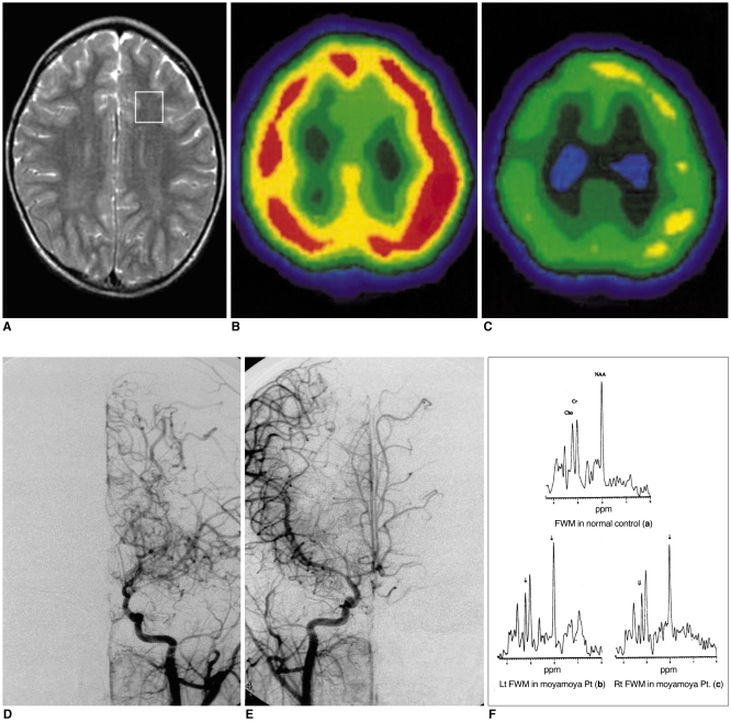
Fig. 1 MRI, basal and diamox stress SPECT, cerebral angiography and 1H-MR spectra obtained in patient No. 5.A. T2-weighted axial image shows a voxel location for 1H-MRS and no abnormal signal intensity in the brain.B. Basal SPECT (axial view) discloses normal uptake in both hemispheres.C. Diamox stress SPECT (axial view) reveals decreased uptake in both hemispheres.D. Arterial-phase angiogram of the left common carotid artery (frontal view) shows marked stenosis of the distal internal carotid and anterior and middle cerebral arteries, with tortuous and dilated medial and lateral lenticulostriate vessels (Suzuki stage II).E. Arterial-phase angiogram of the right common carotid artery (frontal view) demonstrates findings similar to those of the left side (Suzuki stage II).F. Representative 1H-MR spectra of frontal white matter (both sides) show lower NAA/Cr [Lt(b): 1.21, Rt(c): 1.34], Cho/Cr [Lt(b): 0.71, Rt (c): 0.77] ratios than in an age-matched control (a).
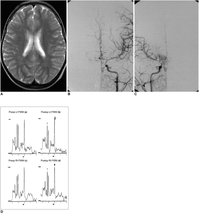
Fig. 2 MRI, cerebral angiography and 1H-MR spectra obtained in patient No. 1.A. T2-weighted axial image shows no abnormal signal intensity in the brain.B. Arterial-phase angiogram of the left common carotid artery (frontal view) reveals narrowing of the carotid fork (Suzuki stage I).C. Angiogram of the right common carotid artery (frontal view) shows the beginning of basal moyamoya vessels, and narrowing of the supraclinoid internal carotid and anterior and middle cerebral arteries (Suzuki stage II).D. 1H-MR spectra of frontal white matter (both sides) obtained after revascularization surgery disclose an increased NAA/Cr ratio [Lt(b): 1.56, Rt(d): 1.41] and a slightly increased upper normal range of Cho/Cr [Lt(b): 1.23, Rt(d): 1.20] as compared with preoperative NAA/Cr [Lt(a): 1.24, Rt(c): 1.14] and Cho/Cr [Lt(a): 0.93, Rt(c): 1.02] levels.
Reference
-
1. Kudoh T. Juvenile occlusion of the circle of Willis. Clin Neurol. 1965; 5:607.2. Kuwabara Y, Ichiya Y, Otsuka M, et al. Cerebral hemodynamic change in the child and the adult with moyamoya disease. Stroke. 1990; 21:272–277. PMID: 2305403.
Article3. Suzuki J, Takaku A. Cerebrovascular "moyamoya" disease: disease showing abnormal net-like vessels in base of brain. Arch Neurol. 1969; 20:288–299. PMID: 5775283.4. Halbach VV, Barkovich AJ. Anomalies of cerebral vasculature: Pediatric neuroimaging. 1995. 2nd ed. New York: Raven Press;p. 619–653.5. Kinugasa K, Mandai S, Kamata I, Sugiu K, Ohmoto T. Surgical treatment of moyamoya disease: operative technique for encephalo-duro-arterio-myo-synangiosis, its follow-up, clinical results, and angiograms. Neurosurgery. 1993; 32:527–531. PMID: 8474642.6. Ishikawa T, Houkin K, Kamiyama H, Abe H. Effects of surgical revascularization on outcome of patients with pediatric moyamoya disease. Stroke. 1997; 28:1170–1173. PMID: 9183345.
Article7. Matsushima Y, Fukai N, Tanaka K, et al. A new surgical treatment of moyamoya disease in children: a preliminary report. Surg Neurol. 1987; 15:313–320. PMID: 7245020.
Article8. Ross BD, Michaelis T. Clinical applications of magnetic resonance spectroscopy. Magn Reson Q. 1994; 10:191–247. PMID: 7873353.9. Kimura H, Fujii Y, Itoh S, et al. Metabolic alterations in neonate and infant brain during development : evaluation with proton MR spectroscopy. Radiology. 1995; 194:483–489. PMID: 7529934.10. Lee JH, Arcinue E, Ross BD. Organic osmolytes in the brain of an infant with hypernatremia. N Engl J Med. 1994; 331:439–442. PMID: 8035840.
Article11. Van der Grond J, Balm R, Kappelle L, Eikelboom BC, Mali WP. Cerebral metabolism of patients with stenosis or occlusion of the internal carotid artery: A 1H-MR spectroscopic imaging study. Stroke. 1995; 26:822–828. PMID: 7740574.12. Kreis R, Ross BD, Farrow NA, Ackerman Z. Metabolic disorders of the brain in chronic hepatic encephalopathy detected with H-1 MR spectroscopy. Radiology. 1992; 182:19–27. PMID: 1345760.
Article13. Bizzi A, Movsas B, Tedeschi G, et al. Response of non-Hodgkin lymphoma to radiation therapy: early and long-term assessment with H-1 MR spectroscopic imaging. Radiology. 1995; 194:271–276. PMID: 7997566.
Article14. Rajanayagam V, Grad J, Krivit W, et al. Proton MR spectroscopy of childhood adrenoleukodystrophy. AJNR Am J Neuroradiol. 1996; 17:1013–1024. PMID: 8791909.15. Duijin JH, Matson GB, Maudsley AA, Hugg JW, Weiner MW. Human brain infarction: Proton MR spectroscopy. Radiology. 1992; 183:711–718. PMID: 1584925.
Article16. Lanfermann H, Kugel H, Heindel W, Herholz K, Heiss W-D, Lackner K. Metabolic changes in acute and subacute cerebral infarctions: findings at proton MR spectroscopic imaging. Radiology. 1995; 196:203–210. PMID: 7784568.
Article17. Van der Grond J, Ramos LMP, Eikelboom BC, Mali WP. Cerebral metabolic differences between the severe and critical hypoperfused brain. Neurology. 1996; 47:399–404. PMID: 8757011.
Article18. Takemichi TK, Prohovnik I, Mohr JP, Correll JW, Quest DO, Jarvis L. Reduced hypercapnic vasoreactivity in moyamoya disease. Neurology. 1988; 38:1575–1581. PMID: 3419602.
Article19. Taki W, Yonekawa Y, Kobayashi A, et al. Cerebral circulation and metabolism in adult's moyamoya disease: PET study. Acta Neurochir (Wien). 1989; 100:150–154. PMID: 2589122.20. Ogawa A, Yoshimoto T, Suzuki J, Sakurai Y. Cerebral blood flow in moyamoya disease, part 1: correlation with age and regional distribution. Acta Neurochir (Wien). 1990; 105:30–34. PMID: 2239376.21. Ogawa A, Nakamura N, Yoshimoto T, Suzuki J. Cerebral blood flow in moyamoya disease, part 2: autoregulation and CO2 response. Acta Neurochir (Wien). 1990; 105:107–111. PMID: 2125802.22. Sato H, Sato N, Tamuki N, Matsumoto S. Chronic low-perfusion state in children with moyamoya disease following revascularization. Childs Nerv Syst. 1990; 6:166–171. PMID: 2357714.
Article23. Ohashi K, Fernandez-Ulloa M, Hall LC. SPECT, magnetic resonance and angiographic features in a moyamoya patient before and after external-to-internal carotid artery bypass. J Nucl Med. 1992; 33:1692–1695. PMID: 1517845.24. Inoue Y, Momose T, Machida K, Honda N, Tsutsumi K. Cerebral vasodilatory capacity mapping using technetium-99m-DTPA-HAS SPECT and acetazolamide in moyamoya disease. J Nucl Med. 1993; 34:1984–1986. PMID: 8229245.25. Rutgers DR, Klijn CJ, Kappelle LJ, van der Grond J. Cerebral metabolic changes in patients with symptomatic occlusion of the internal carotid artery: a longitudinal 1H magnetic resonance spectroscopy study. J Magn Reson Imaging. 2000; 11:279–286. PMID: 10739559.26. Janson C, McPhee S, Bilaniuk L, et al. Clinical protocol gene therapy of Canavan disease: AAV-2 vector for neurosurgical delivery of aspartoacylase gene (ASPA) to the human brain. Hum Gene Ther. 2002; 13:1391–1412. PMID: 12162821.27. Hashimoto T, Tayama M, Miyazaki M, et al. Developmental brain changes investigated with proton magnetic resonance spectroscopy. Dev Med Child Neurol. 1995; 37:398–405. PMID: 7768339.
Article28. Shimizu H, Shirane R, Fujiware S, Takahashi A, Yoshimoto T. Proton magnetic resonance spectroscopy in children with moyamoya disease. Clin Neurol Neurosurg. 1997; 99:S64–S67. PMID: 9409409.
Article29. Touho H, Karasawa J, Ohnishi H. Preoperative and postoperative evaluation of cerebral perfusion and vasodilatory capacity with 99mTc-HMPAO SPECT and acetazolamide in childhood moyamoya disease. Stroke. 1996; 27:282–289. PMID: 8571424.30. Hoshi H, Ohnishi T, Jinnouchi S, et al. Cerebral blood flow study in patients with moyamoya disease evaluated by IMP SPECT. J Nucl Med. 1994; 35:44–50. PMID: 8271059.
- Full Text Links
-
- Actions
-
Cited
- CITED
-
- Close
- Share
-
- Similar articles
-
- Neuroimaging Diagnosis and Treatment of Moyamoya Disease
- Localized, water-suppressed in vivo H MR spectroscopy of human brain tumors: Preliminary results
- In vivo H MR spectroscopy of human brain in six normal volunteers
- Metabolic Changes after Revascularization in a Patient with Innominate Artery Occlusion by Localized in vivo Proton Magnetic Resonance Spectroscopy
- Usefulness of 1H Magnetic Resonance Spectroscopy for Evaluation of Cognitive Impairment after Subarachnoid Hemorrhage Caused by Rupture of Middle Cerebral Artery Aneurysm
