Korean J Ophthalmol.
2004 Jun;18(1):47-51. 10.3341/kjo.2004.18.1.47.
Diplopia and Periorbital Mass Associated with Miragel Buckling Explant
- Affiliations
-
- 1The Institute of Vision Research, College of Medicine, Yonsei University, Seoul, Korea.
- 2Department of Ophthalmology, College of Medicine, Yonsei University, Seoul, Korea.
- KMID: 1115783
- DOI: http://doi.org/10.3341/kjo.2004.18.1.47
Abstract
- A 28-year-old female presented with a palpable mass lesion on the superonasal aspect of her right globe and she had a progressive diplopia. She had a scleral encircling surgery with a Miragel explant (MIRA, Waltham, Mass, USA) for the tractional retinal detachment associated with pars planitis 9 years previously. On examination, she revealed restricted eye movements of her right eye. The magnetic resonance imaging documented a swelling of the Miragel explant that mimicked a periorbital mass lesion. The Miragel explant was removed and fragmentation of the explant was found intraoperatively. The removed Miragel explant was examined by a scanning electron microscopy, and this demonstrated a disintergrated and swollen structural composition of the Miragel explant. Postoperatively, her extraocular movement was almost restored and the retina remained well attached. Alterations in the structural composition of the Miragel explant results in an excessive swelling that causes a restriction of the extraocular movement, and this can mimick a periorbital mass lesion.
MeSH Terms
Figure
-
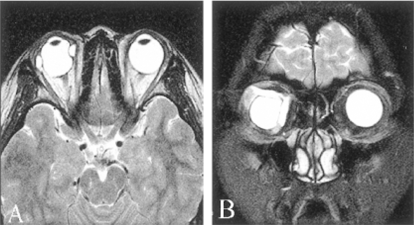
Fig. 1A,B The magnetic resonance imaging documents the enlargement of the encircling Miragel explant. Sagittal view (A), Coronal view (B).

Fig. 2 A preoperative Hess screen test showing a severely restricted extaocular movement in her right eye (A). A postoperative Hess screen test showing the marked improvement in eye movements (B).
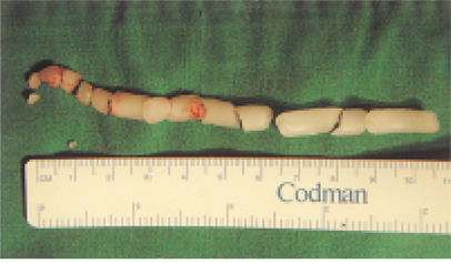
Fig. 3 The Miragel explant shows extreme friability and a tendency to disintegrate into fragments intraoperatively.
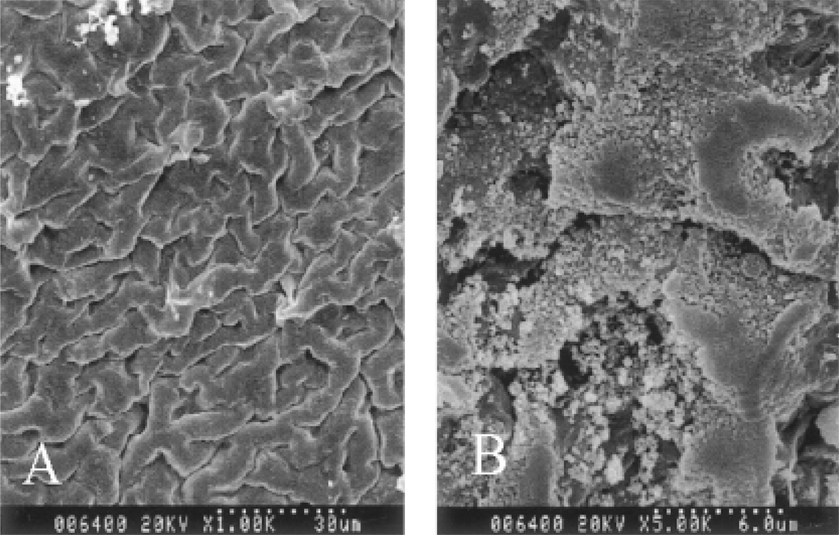
Fig. 4A,B The removed Miragel material demonstrated a structural deterioration with micropores, a distorted shape and an irregularity in size upon scanning electron microscopy (A: × 1000, B: × 5000).
Reference
-
1. Ho PC, Chan IM, Refojo MF, Tolentino FI. The MAI hydrophilic implant for scleral buckling: a review. Ophthalmic Surg. 1984. 15:511–515.2. Marin JF, Tolentino FI, Refojo MF, Schepens CL. Long-term complications of the MAI hydrogel intrascleral buckling implant. Arch Ophthalmol. 1992. 110:86–88.3. Rolden-Pallares M, del Castillo Sanz JL, Awad-El Susi S, Refojo MF. Long-term complications of silicone and hydrogel explants in retinal reattachment surgery. Arch Ophthalmol. 1999. 117:197–201.4. Hwang KI, Lim JL. Hydrogel explant fragmentation 10 years after scleral buckling surgery. Arch Ophthalmol. 1997. 115:1205–1206.5. Braunstein RA, Winnick M. Complications of Miragel: Pseudotumor. Arch Ophthalmol. 2002. 120:228–229.6. Li K, Lim KS, Wong D. Miragel explant fragmentation 10 years after scleral buckling surgery. Eye. 2003. 17:248–250.7. D'Hermies F, Korobelnik JF, Savoldelli M, Chauvaud D, Pouliquen Y. Miragel versus silastic used as episcleral implants in rabbits. Retina. 1995. 15:62–67.8. D'Hermies F, Korobelnik JF, Caputo G, Mashhour B, Chauvaud D, Pouliquen Y, Renard G. Encapsulation of scleral buckling materials A studyof sixty specimens. Ophthalmology. 1998. 105:1079–1086.9. Le Rouic JF, Bejjani RA, Azan F, Bettembourg O, Renard G, Chauvaud D. Cryoextraction of episcleral Miragel buckle elements: a new technique to reduce fragmentation. Ophthalmic Surg Lasers. 2002. 33:237–239.10. Refojo MF, Natchiar G, Liu HS, Lahav M, Tolentino FI. New hydrophilic implant for scleral buckling. Ann Ophthalmol. 1980. 12:88–92.11. Refojo MF, Leong FL. Poly(methyl acrlate-co-hydrocyethly acrylate) hydrogel implant material of strength and softness. J Biomed Mater Res. 1981. 15:497–509.12. Tolentino FI, Refojo MF, Schepens CL. A hydrophilic acrylate implant for scleral buckling: technique and clinical experience. Retina. 1981. 1:281–286.
- Full Text Links
-
- Actions
-
Cited
- CITED
-
- Close
- Share
-
- Similar articles
-
- Late Complications after Successful Scleral Buckling Surgery Using Hydrogel Buckles
- Ocular Movement Disorder after Scleral Buckling Surgery in Patients with Retinal Detachment
- Subconjunctival Abscess Formation with Periorbital Cellulitis Following Scleral Buckling
- 2 Cases of Combined Cataract and Scleral Buckling Surgery
- A Case of Pneumocele in the Ethmoid Sinus
