Spontaneous Regression of Neovascularization at the Disc in Diabetic Retinopathy
- Affiliations
-
- 1Department of Ophthalmology, Hallym University College of Medicine, Hallym University Sacred Heart Hospital, Anyang, Korea.
- KMID: 1115782
- DOI: http://doi.org/10.3341/kjo.2004.18.1.41
Abstract
- Neovascularization at the disc (NVD) is the most serious complication in diabetic retinopathy, and leads to vitreous hemorrhage and tractional retinal detachment. We report two cases of spontaneous regression of NVD in proliferative diabetic retinopathy. Two men (31 and 46 years old) with diabetes had NVD in both eyes. They were treated with panretinal photocoagulation on the left eye first, but their right eyes went untreated, because they did not revisit our clinic for several months. Fortunately, on revisit, their neovascularization had disappeared a few months later in both eyes, including their untreated right eyes. We could not find any specific causes for the spontaneous regression of the new vessels.
MeSH Terms
Figure
-
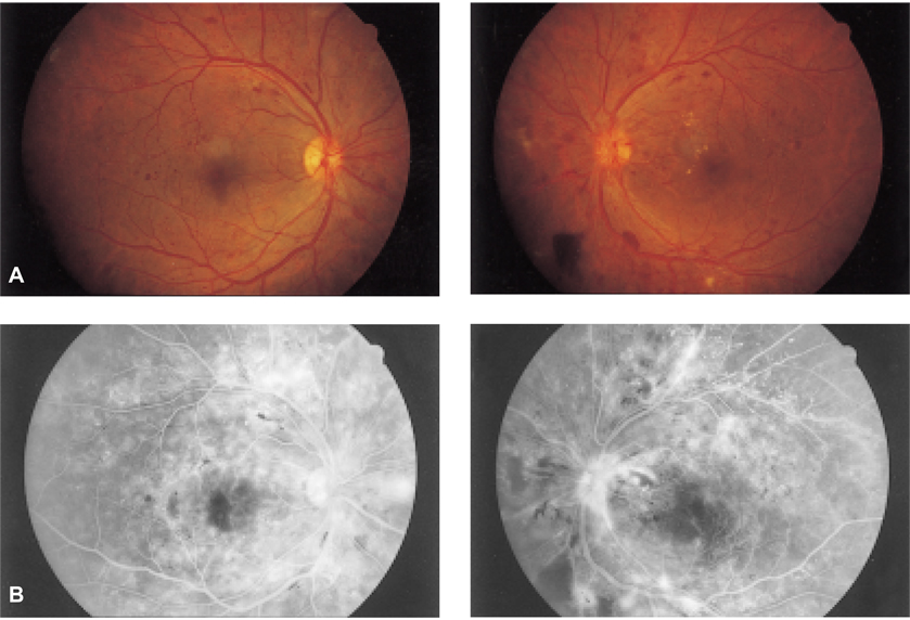
Fig. 1 Case 1. (A) Top: With proliferative DM retinopathy, neovascularization of the optic disc is seen on both eyes. (B) Bottom: Angiography shows multiple, severe leakage from neovascularization of the disc and retina on both eyes.

Fig. 2 Case 1. (A) Top: Neovascularization of the optic disc has disappeared on both eyes. (B) Bottom: Angiography shows no active leakage at the disc on either eyes.
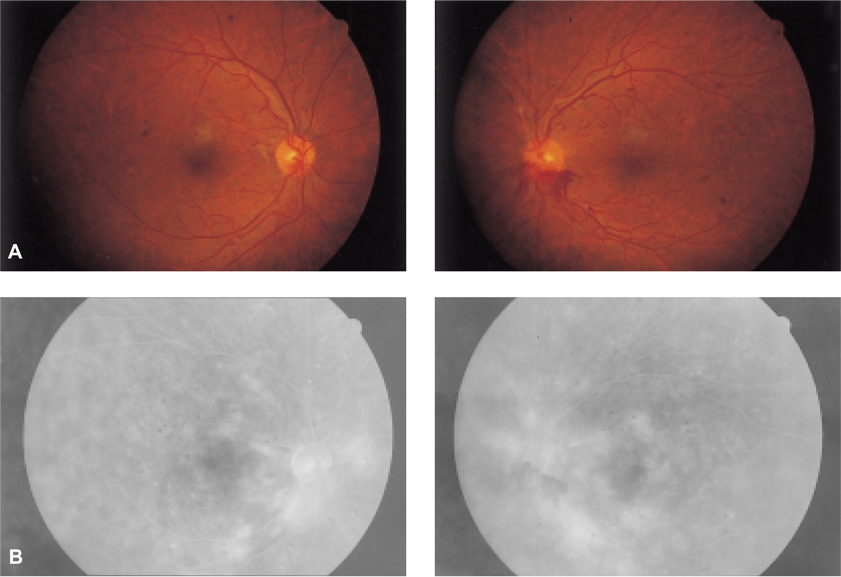
Fig. 3 Case 2. (A) Top: With proliferative DM retinopathy, neovascularization of the optic disc is seen on both eyes. (B) Bottom: Angiography shows leakage from neovascularization of the disc and retina on both eyes.
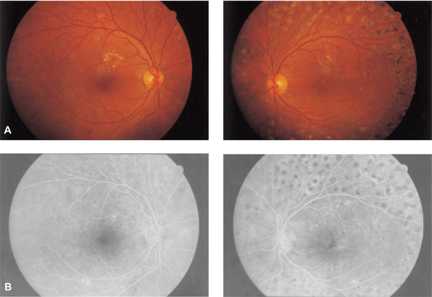
Fig. 4 Case 2. (A) Top: neovascularization of the optic disc has disappeared and some hard exudates are seen at the posterior pole on both eyes. (B) Bottom: Angiography shows no active leakage at the disc on either eyes.
Reference
-
1. Bandello F, Gass JD, Lattanzio R, Brancato R. Spontaneous regression of neovascularization at the disk and elsewhere in diabetic retinopathy. Am J Ophthalmol. 1996. 122:494–501.2. Henkind P. Ocular neovascularization. Am J Ophthalmol. 1978. 85:287–301.3. Patz A. Clinical and experimental studies on retinal neovascularization. Am J Ophthalmol. 1982. 94:715–743.4. Yaval Y, Linda WP, Stuart LF, Lawrence S, David HO, Arnall P. Optic disc neovascularisation in diabetic retinopathy: II. Natural history and results of photocoagulation treatment. Br J Ophthalmol. 1980. 64:77–86.5. Taylor E, Dobree JH. Proliferative diabetic retinopathy: site and size of initial lesions. Br J Ophthalmol. 1970. 54:11–23.6. Little HL. Argon laser photocoagulation of proliferative diabetic retinopathy. Int Ophthalmol Clin. 1976. 16:79–103.7. Diabetic Retinopathy Study Research Group. Photocoagulation treatment of proliferative diabetic retinopathy: the second report of Diabetic Retinopathy Study findings. Ophthalmology. 1978. 85:82–106.8. Shorb SR, Irvine AR, Kimura SJ, Morris BW. Optic disk neovascularization associated with chronic uveitis. Am J Ophthalmol. 1976. 82:175–182.9. Schatz H, Patz A. Cystoid maculopathy in diabetics. Arch Ophthalmol. 1976. 94:761–768.10. Kearns M, Hamilton AM, Kohner EM. Excessive permeability in diabetic maculopathy. Br J Ophthalmol. 1979. 63:489–497.11. Bonnet M, Bensoussan B, Grange JD, Pingault C, Francoz N. Capillaropathie oedemateuse aigue du diabetique insulin-dependant. J Fr Ophtalmol. 1982. 5:303–316.12. Beetham WP. Visual prognosis of proliferative diabetic retinopathy. Br J Ophthalmol. 1963. 47:611–619.13. Davis MD. Vitreous contraction in proliferative diabetic retinopathy. Arch Ophthalmol. 1965. 74:741–751.14. Dobree JH. Proliferative diabetic retinopathy: evolution of the retinal lesions. Br J Ophthalmol. 1964. 48:637–649.15. Ramsay WJ, Ramsay RC, Purple RL, Knoblock WH. Involutional diabetic retinopathy. Am J Ophthalmol. 1977. 84:851–858.16. Davis MD. Ryan SJ, editor. Proliferative diabetic retinopathy. Retina. 1989. St. Louis: CV Mosby Co.;367–402.17. Brancato R, Menchini U, Brndello F. Diabetic papillopathy: fluoroangiographic aspects. Metab Pediatr Syst Ophthalmol. 1986. 9:57–61.18. Peter JK, John JW. Resolution of optic disk neovascularization associated with intraocular inflammation. Am J Ophthalmol. 1980. 90:545–548.19. Shabo AL, Maxwell DS. Experimental immunogenic proliferative retinopathy in monkeys. Am J Ophthalmol. 1977. 83:471–480.20. Brucker AJ. Disk and peripheral retina neovascularization secondary to talc and cornstarch emboli. Am J Ophthalmol. 1979. 88:864–868.21. Foos RY. Vitreoretinal juncture, topographical variations. Invest Ophthalmol. 1972. 11:801–806.22. Asdourian GK, Goldberg MF, Busse B. Optic disk neovascularization of uveal origin. Arch Ophthalmol. 1977. 95:998–1002.
- Full Text Links
-
- Actions
-
Cited
- CITED
-
- Close
- Share
-
- Similar articles
-
- Two Cases of Choroidal Neovascularizatien After Photocoagulation
- Spontaneous Regression of a Large Lumbar Disc Extrusion
- Comparison of venous filling times and SLO findings at each quadrant region in diabetic retinopathy
- The Effects of Argon Laser Photocoagulation in Diabetic Retinopathy
- Clinical Study on The Vitreous Hemorrhage
