A Case of Accidental Macular Injury by Nd: YAG Laser and Subsequent 6 Year Follow-Up
- Affiliations
-
- 1Department of Ophthalmology, School of Medicine, Kyungpook National University, Daegu, Korea. itkim@knu.ac.kr
- KMID: 1115755
- DOI: http://doi.org/10.3341/kjo.2009.23.3.207
Abstract
- Here, we report the case of a patient who sustained Nd: YAG laser macular injury with subsequent 6 year follow-up evaluation. A 23-year-old female was accidentally exposed to a Q-switched Nd: YAG laser without protective goggles. Upon initial evaluation, the best-corrected visual acuity of her affected eye was 20/100 OD. Fundoscopic examination revealed a macular laser burn and vitreous hemorrhage. Corticosteroids, in the form of 60 mg prednisolone, were administered orally with a 10 mg per week taper. Nineteen days following exposure, fundoscopic examination revealed a distinct epiretinal membrane which resolved within six months. The best-corrected visual acuity of the affected eye remained 20/100 OD. This clinical course is similar to those of previously reported cases including vitreous hemorrhage and subsequent epiretinal membrane formation. However, visual acuity did not recover despite spontaneous regression of the epiretinal membrane and at 6 year follow-up, there was neither choroidal neovascularization nor macular hole formation.
MeSH Terms
-
Accidents
Female
Fluorescein Angiography
Follow-Up Studies
Fundus Oculi
Glucocorticoids/therapeutic use
Humans
Lasers, Solid-State/*adverse effects
Macula Lutea/*injuries
Prednisolone/therapeutic use
*Radiation Injuries/complications/diagnosis/drug therapy/physiopathology
Treatment Outcome
Visual Acuity/radiation effects
Vitreous Hemorrhage/etiology/pathology
Young Adult
Figure
-

Fig. 1 Fundoscopic photograph on the day of injury revealed vitreous hemorrhage and the site of laser injury at the macula.
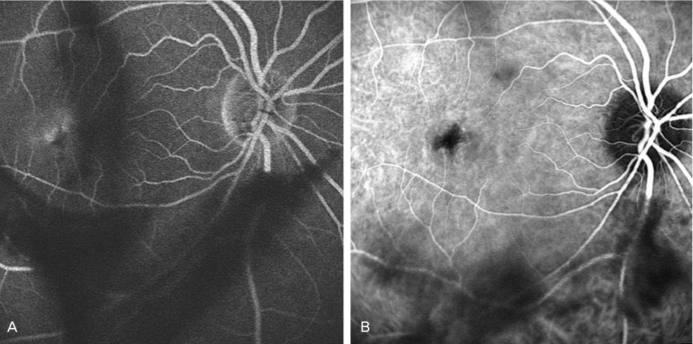
Fig. 2 (a) One day following the exposure, fluorescein angiography revealed a round area of hypofluorescence with late macular hyperfluorescence. (b) Two days later, indocyanine green angiography failed to demonstrate hyperfluorescence.
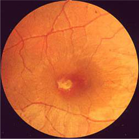
Fig. 3 Nineteen days following the exposure, fundoscopic photography revealed an epiretinal membrane with retinal folds radiating to the parafoveal area.
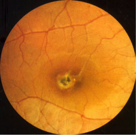
Fig. 4 Six months later, fundoscopic photography revealed spontaneous regression of the epiretinal membrane and central pigmentation of the laser burn site.

Fig. 5 Six years later, fundoscopic photography revealed a chorioretinal scar with more prominent central pigmentation.
Reference
-
1. Boldrey EE, Little HL, Flocks M, Vassiliadis A. Retinal injury due to industrial laser burns. Ophthalmology. 1981. 88:101–107.2. Kearney JJ, Cohen HB, Stuck BE, et al. Laser injury to multiple retinal foci. Lasers Surg Med. 1987. 7:499–502.3. Glovinsky Y, Regenbogen L, Bartov E, et al. Macular pucker following accidental laser burn. Metab Pediatr Syst Ophthalmol. 1982. 6:355–359.4. Thach AB, Lopez PF, Snady-McCoy LC, et al. Accidental Nd: YAG laser injuries to the macula. Am J Ophthalmol. 1995. 119:767–773.5. Lam TT, Tso MO. Retinal injury by neodymium: YAG laser. Retina. 1996. 16:42–46.6. Ray S, Topping T, Young LH. Spontaneous peeling of epiretinal membrane associated with Nd:YAG laser injury. Arch Ophthalmol. 2001. 119:137–139.7. Asano T. Accidental YAG laser burn. Am J Ophthalmol. 1984. 98:116–117.8. Ryan SJ. Subretinal neovascularization. Natural history of an experimental model. Arch Ophthalmol. 1982. 100:1804–1809.9. Wolfe JA. Laser retinal injury. Mil Med. 1985. 150:177–185.10. Gabel VP, Birngruber R, Lorenz B, Lang GK. Clinical observations of six cases of laser injury to the eye. Health Phys. 1989. 56:705–710.
