MR Imaging Findings of Ovarian Cystadenofibroma and Cystadenocarcinofibroma: Clues for the Differential Diagnosis
- Affiliations
-
- 1Department of Radiology, Seoul National University College of Medicine, Seoul, Korea. kimsh@radcom.snu.ac.kr
- KMID: 1114353
- DOI: http://doi.org/10.3348/kjr.2006.7.3.199
Abstract
OBJECTIVE
We wanted to assess the MR imaging findings of ovarian cystadenofibroma and cystadenocarcinofibroma, and we wanted to find clues for making the differential diagnosis between them. MATERIALS AND METHODS: The MR images of 12 pathologically proven cystadenofibromas and two cystadenocarcinofibromas were reviewed, with a focus on the internal architecture, signal intensity and enhancement. RESULTS: All the tumors appeared as multilocular cysts, except for a single unilocular cystic mass and a single solid mass. The previously reported characteristic MR findings of cystadenofibroma (a multilocular cystic mass with a T2-dark-signal-intensity solid component containing small cystic locules) were found in only 43% of the tumors (6/14). Diffuse or partial thickening of the cyst wall with T2-dark signal intensity without a definite solid component was as common as the previous reported findings (6/14). Two cystadenocarcinofibromas showed more prominent solid portions with higher T2-signal intensities and stronger enhancement than did the cystadenofibromas. CONCLUSION: Diffuse or partial thickening of the cyst wall with dark-signal-intensity in multilocular cystic masses may suggest ovarian cystadenofibroma, and this type of appearance may be as common as the previously reported characteristic appearance. A prominent solid component with a higher T2-signal intensity and strong enhancement are the typical findings of cystadenocarcinofibroma.
Keyword
MeSH Terms
Figure
-

Fig. 1 A 42-year-old woman with a mucinous cystadenofibroma. The T1- (A) and T2-weighted (B) MR images show a multilocular cystic mass with a solid component (arrows) in the right ovary. The solid component (black arrowheads) shows low signal intensity and it contains multiple, small cystic locules (white arrowheads). C. The contrast-enhanced T1-weighted sagittal MR image with fat-suppression shows mild enhancement in the solid component (arrowheads) within mass (arrows).

Fig. 2 A 24-year-old woman with a mucinous cystadenofibroma. A. The T2-weighted sagittal MR image shows a huge multilocular cystic mass (arrows) with a solid component in the left ovary. The solid component with low signal intensity contains small cystic locules (arrowheads). B. The contrast-enhanced T1-weighted sagittal MR image with fat-suppression shows mild enhancement in the solid component (arrowheads) within mass (arrows).

Fig. 3 A 58-year-old woman with a serous cystadenofibroma. The T1- (A) and T2-weighted (B) MR images show a multilocular cystic mass in the right adnexal region (arrows). The wall of the mass was irregularly and partially thickened, and it appeared as dark signal intensity on this T2-weighted image.

Fig. 4 A 58-year-old woman with a serous cystadenofibroma. The T2-weighted MR images in the axial (A) and sagittal (B) planes show a bilocular cystic mass (arrows) surrounded by a partly-thickened wall of dark-signal-intensity (small arrows) in the left ovary. Note the presence of focal nodular thickening (arrowheads).

Fig. 5 A 62-year-old woman with an endometrioid cystadenocarcinofibroma. A. The T2-weighted MR image in the sagittal plane shows a multilocular cystic mass (arrows) with a solid component (black arrowheads) in the left ovary. Partial thickening with dark-signal-intensity was noted in the superior part of the wall (white arrowheads). The solid component shows relatively higher signal intensity compared with the benign counterpart in Figure 1 and 2. B. The contrast-enhanced T1-weighted sagittal MR image with fat-suppression shows strong enhancement in the mass (arrows).
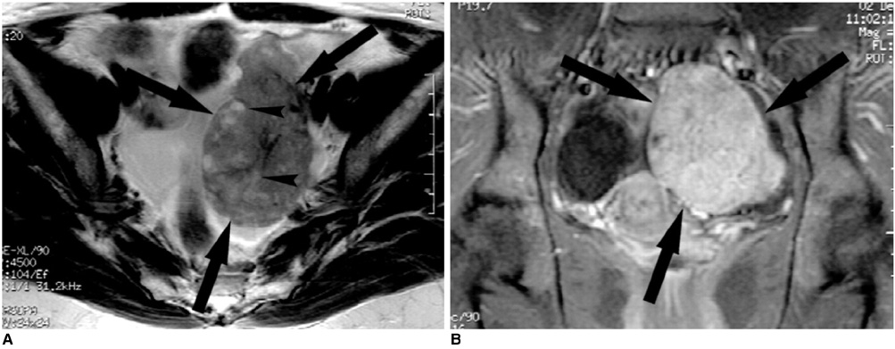
Fig. 6 A 43-year-old woman with serous cystadenocarcinofibroma. A. The T2-weighted MR image shows a mainly solid mass (arrows) in the left ovary. The mass contained multiple, small cystic locules (arrowheads) and it was of intermediate signal intensity. B. The contrast-enhanced T1-weighted MR image shows strong enhancement in the solid component (arrows).
Reference
-
1. Czernobilsky B, Borenstein R, Lancet M. Cystadenofibroma of the ovary: a clinicopathologic study of 34 cases and comparison with serous cystadenoma. Cancer. 1974. 34:1971–1981.2. Kao GF, Norris HJ. Cystadenofibroma of the ovary with epithelial atypia. Am J Surg Pathol. 1978. 2:357–363.3. Groutz A, Wolman I, Wolf Y, Luxman D, Sagi J, Jaffa AJ, et al. Cystadenofibroma of the ovary in young women. Eur J Obstet Gynecol Reprod Biol. 1994. 54:137–139.4. Fatum M, Rojansky N, Shushan A. Papillary serous cystadenofibroma of the ovary -- is it really so rare? Int J Gynaecol Obstet. 2001. 75:85–86.5. Outwater EK, Siegelman ES, Talerman A, Dunton C. Ovarian fibromas and cystadenofibromas: MRI features of the fibrous component. J Magn Reson Imaging. 1997. 7:465–471.6. Takeuchi M, Matsuzaki K, Kusaka M, Shimazu H, Yoshida S, Nishitani H, et al. Ovarian cystadenofibromas: characteristic magnetic resonance findings with pathologic correlation. J Comput Assist Tomogr. 2003. 27:871–873.7. Cho SM, Byun JY, Rha SE, Jung SE, Park GS, Kim BK, et al. CT and MRI findings of cystadenofibromas of the ovary. Eur Radiol. 2004. 14:798–804.8. Gougoutas CA, Siegelman ES, Hunt J, Outwater EK. Pelvic endometriosis: various manifestations and MR imaging findings. AJR Am J Roentgenol. 2000. 175:353–358.9. Sala EJ, Atri M. Magnetic resonance imaging of benign adnexal disease. Top Magn Reson Imaging. 2003. 14:305–327.10. Jung SE, Lee JM, Rha SE, Byun JY, Jung JI, Hahn ST. CT and MR imaging of ovarian tumors with emphasis on differential diagnosis. Radiographics. 2002. 22:1305–1325.11. Kim KA, Park CM, Lee JH, Kim HK, Cho SM, Kim B, et al. Benign ovarian tumors with solid and cystic components that mimic malignancy. AJR Am J Roentgenol. 2004. 182:1259–1265.12. Moon WJ, Koh BH, Kim SK, Kim YS, Rhim HC, Cho OK, et al. Brenner tumor of the ovary: CT and MR findings. J Comput Assist Tomogr. 2000. 24:72–76.13. Kim SH, Kim WH, Park KJ, Lee JK, Kim JS. CT and MR findings of Krukenberg tumors: comparison with primary ovarian tumors. J Comput Assist Tomogr. 1996. 20:393–398.14. Joja I, Asakawa T, Mitsumori A, Nakagawa T, Hiraki Y, Kudo T, et al. Struma ovarii: appearance on MR images. Abdom Imaging. 1998. 23:652–656.15. Stevens SK, Hricak H, Stern JL. Ovarian lesions: detection and characterization with gadolinium enhanced MR imaging at 1.5 T. Radiology. 1991. 181:481–488.
- Full Text Links
-
- Actions
-
Cited
- CITED
-
- Close
- Share
-
- Similar articles
-
- MR Imaging Findings of Ovarian Cystadenofibroma: Clues for Making the Differential Diagnosis from Ovarian Malignancy
- A Case of Cystadenofibroma of the Ovary
- Pearls and Potential Pitfalls for Correct Diagnosis of Ovarian Cystadenofibroma in MRI: A Pictorial Essay
- Sonographic Features of Ovarian Fibromatous Tumors: Atypical Cystic Lesion
- A clear cell cystadenofibroma of the ovary
