Prenatal Sonographic and MR Imaging Findings of Extensive Fetal Lymphangioma: A Case Report
- Affiliations
-
- 1Department of 1Radiology, The Catholic University of Korea, College of Medicine. jybyun@catholic.ac.kr
- 2Department of Radiology, University of Ulsan College of Medicine.
- 3Department of Obstetrics and Gynecology, The Catholic University of Korea, College of Medicine.
- 4Department of Pathology, The Catholic University of Korea, College of Medicine.
- KMID: 1111257
- DOI: http://doi.org/10.3348/kjr.2003.4.4.260
Abstract
- We report the imaging findings in a case of fetal lymphangioma involving the retroperitoneum and right lower extremity, and diagnosed by ultrasonography and magnetic resonance (MR) imaging at 26 weeks of gestation. Prenatal ultrasonograms and T2-weighted single-shot fast spin-echo MR images clearly revealed an extensive, multilocular cystic mass with internal hemorrhage in the retroperitoneum extending to the lower extremity.
Keyword
MeSH Terms
Figure
-
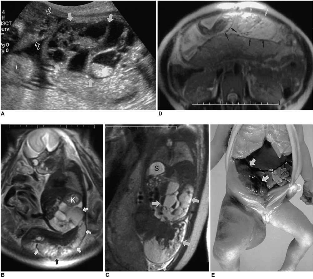
Fig. 1 A 26-week-old female fetus with an extensive lymphangioma. A. Prenatal ultrasonography shows a large multiseptated cystic mass (arrows) with variable echo patterns, located at the right of the fetal abdomen and also involving the right lower extremity (open arrows). The transverse diameter of this extremity is much greater than that of the left one (L). Color Doppler ultrasonography revealed no flow within the mass at the time of examination (not shown). B. Sagittal T2-weighted single-shot fast spin-echo MR image (TR/TE, 1624/79) depicts a large intra-abdominal high-signal-intensity mass (arrows) with multiple internal locules, displacing the ipsilateral kidney (K) superiorly. The mass (arrows) extends continuously to the right buttock and right lower extremity. C. Coronal T2-weighted single-shot fast spin-echo MR image (TR/TE, 1624/79) clearly reveals the multilocular cystic nature of the mass (arrows), each locule of which contains multiple fluid-fluid levels. S=Stomach. D. T2-weighted single-shot fast spin-echo MR image (TR/TE, 1624/79) obtained parallel to the right lower fetal extremity shows the distal extent of the lymphangioma (arrows). This presents as an extensive subcutaneous cystic mass, with asymmetric limb hypertrophy. E. Post-mortem photograph depicts a large lymphangioma (arrows) at the right of the abdomen, extending to the right lower extremity.
Reference
-
1. Marches C, Savin E, Dragone E, et al. Cystic hygroma: prenatal diagnosis and genetic counseling. Prenat Diagn. 1985. 5:221–227.2. Deshpande P, Twining P, O'Neill D. Prenatal diagnosis of fetal abdominal lymphangioma by ultrasonography. Ultrasound Obstet Gynecol. 2001. 17:445–448.3. Suzuki N, Tsuchida Y, Takahashi A, et al. Prenatally diagnosed cystic lymphangioma in infants. J Pediatr Surg. 1998. 33:1599–1604.4. Kaminopetros P, Jauniaux E, Kane P, Weston M, Nicolaides KH, Campbell DJ. Prenatal diagnosis of an extensive fetal lymphangioma using ultrasonography, magnetic imaging and cytology. Br J Radiol. 1997. 70:750–753.5. Breysem L, Bosmans H, Dymarkowski S, et al. The value of fast MR imaging as an adjunct to ultrasound in prenatal diagnosis. Eur Radiol. 2003. 13:1538–1548.6. Kosir MA, Sonnino RE, Gauderer MW. Pediatric abdominal lymphangiomas: a plea for early recognition. J Pediatr Surg. 1991. 26:1309–1313.7. Ho M, Lee CC, Lin TY. Prenatal diagnosis of abdominal lymphangioma. Ultrasound Obstet Gynecol. 2002. 20:203–208.8. Romero R, Pilu G, Jeanty P, Ghidini A, Hobbins JC. Romero R, Pilu G, Jeanty P, Ghidini A, Hobbins JC, editors. Cystic hygroma. Prenatal diagnosis of congenital anomalies. 1988. Norwalk, CN: Appleton & Lange;115–118.9. Lee SH, Cho JY, Song MJ, et al. Prenatal ultrasound findings of fetal neoplasms. Korean J Radiol. 2002. 3:64–73.10. Kubik-Huch RA, Huisman TAG, Wisser J, et al. Ultrafast MR imaging of the fetus. AJR Am J Roentgenol. 2000. 174:1599–1606.11. Dubois J, Garel L, Abela A, Laberge L, Yazbeck S. Lymphangiomas in children: percutaneous sclerotherapy with an alcoholic solution of zein. Radiology. 1997. 204:651–654.12. Watari H, Yamada H, Fujino T, et al. A case of intrauterine medical treatment for cystic hygroma. Eur J Obstet Gynecol Reprod Biol. 1996. 70:201–203.
- Full Text Links
-
- Actions
-
Cited
- CITED
-
- Close
- Share
-
- Similar articles
-
- Fetal Axillary Cystic Lymphangioma Detected by Prenatal Ultrasonography
- A Case of Prenatally Diagnosed Fetal Retroperitoneal Cystic Lymphangioma
- Prenatal ultrasonographic findings of a case of multiple pterygium syndrome
- Antenatal Diagnosis of Iniencephaly: Sonographic and MR Correlation: A Case Report
- Prenatal MRI Findings of Polycystic Kidney Disease Associated with Holoprosencephaly
