Prenatal Sonographic Diagnosis of Focal Musculoskeletal Anomalies
- Affiliations
-
- 1Department of Diagnostic Radiology, Samsung Cheil Hospital, Sungkyunkwan University School of Medicine. radjycho@skku.edu
- 2Department of Diagnostic Pathology, Samsung Cheil Hospital, Sungkyunkwan University School of Medicine.
- KMID: 1111254
- DOI: http://doi.org/10.3348/kjr.2003.4.4.243
Abstract
- Focal musculoskeletal anomalies vary, and can manifest as part of a syndrome or be accompanied by numerous other conditions such as genetic disorders, karyotype abnormalities, central nervous system anomalies and other skeletal anomalies. Isolated focal musculoskeletal anomaly does, however, also occur; its early prenatal diagnosis is important in deciding prenatal care, and also helps in counseling parents about the postnatal effects of numerous possible associated anomalies. We have encountered 50 cases involving focal musculoskeletal anomalies, including focal limb dysplasia [radial ray abnormality (n=3), mesomelic dysplasia (n=1) ]; anomalies of the hand [polydactyly (n=8), syndactyly (n=3), ectrodactyly (n=1), clinodactyly (n=6), clenched hand (n=5) ]; anomalies of the foot [clubfoot (n=10), rockerbottom foot (n=5), sandal gap deformity (n=1), curly toe (n=2) ]; amniotic band syndrome (n=3) ; and anomalies of the focal spine [block vertebra (n=1), hemivertebra (n=1) ]. Among these 50 cases, five [polydactyly (n=1), syndactyly (n=2) and curly toe (n=2) ] were confirmed by postnatal physical evaluation, two (focal spine anomalies) were diagnosed after postnatal radiologic examination, and the remaining 43 were proven at autopsy. For each condition, we describe the prenatal sonographic findings, and include a brief review.
MeSH Terms
Figure
-

Fig. 1 Radial ray abnormality. Second-trimester ultrasound reveals that the forearm consists of only the ulna (arrow), with sharp radial deviation of the hand. The thumb is not well visualized, consistent with radial ray abnormality.

Fig. 2 Focal skeletal dysplasia: mesomelia. Fetal sonography performed at week 20 demonstrates the typical finding of mesomelic dysplasia. The forearm (radius and ulnar, arrows) is markedly shortened compared with the humerus.
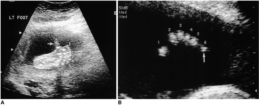
Fig. 3 Polydactyly. A. Prenatal ultrasound reveals that the foot, viewed radially, has an extra digit (preaxial polydactyly) (arrow). B. An extra digit at the ulnar side of hand, viewed axially, suggests postaxial polydactyly (arrow).
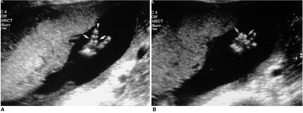
Fig. 4 Syndactyly. A, B. At prenatal sonography, two digits (arrows) at the ulnar side of a fetal hand are seen to be stuck together. The distal phalanges of these digits are observed as one bony segment (arrowhead).

Fig. 5 Clenched hand deformity. At fetal ultrasound, overlapping of the fourth and fifth fingers radially, and the second finger in an ulnar direction, are observed consistently in a fetus with trisomy 18.

Fig. 6 Ectrodactyly (split hands or feet). A. The hand of this fetus has only four fingers, with abnormal widening between the second and third finger (arrows). B. Neonatal photograph demonstrates lobster claw deformity of the hand.

Fig. 7 Clinodactyly. Second trimester ultrasound reveals shortening and radial deviation of the middle phalanx of the fifth finger (arrow), a condition frequently associated with karyotype abnormality such as trisomy 21,18 and 13.

Fig. 8 Clubfoot. At 22 second weeks' gestation, the forefoot (arrows) is oriented in the same plane as the lower leg.
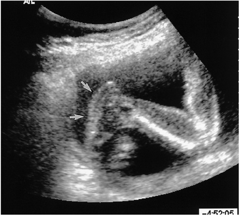
Fig. 9 Rockerbottom foot. Prenatal ultrasound demonstrates eversion of the plantar arch (arrows).
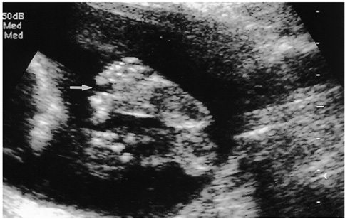
Fig. 10 Sandal gap deformity. Plantar view of the foot in a fetus with Down syndrome reveals an abnormal degree of separation between the great toe and the second toe (arrow).

Fig. 11 Curly toe. A, B. Prenatal sonography indicates that the fourth toe (arrow) deviates medially and plantarly (A, axial scan; B, coronal scan). C. Neonatal photograph confirms the presence of a congenital underlapping fourth toe (arrow).

Fig. 12 Amniotic band syndrome. A. In this fetus with ABS, focal constriction (arrows) at the ankle has induced swelling of the foot. B. Amniotic bands are seen in the vicinity of the fetal hand (arrow), explaining the presence of focal lymphedema in the distal hand. C. In another fetus with ABS, the left arm is amputated at the mid portion of the humerus (arrows). D. Radiograph of the autopsy specimen in related to figure C confirms amputation below mid-humerus level (arrow).

Fig. 13 Hemivertebra and block vertebra. A, B. Hypoplastic right vertebral body at the low lumbar level results in a focal defect at sagittal (A) and axial (B) scanning (arrow), diagnosed as hemivertebra. C. Oblique coronal prenatal ultrasound scan obtained at the low lumbar level suggests the fusion of two vertebral bodies, leading to an abnormally elongated vertebral column compared with other normal body contours. D. Postnatal plain film of the fetus in Fig. 12C demonstrates block vertebra at the right side between the fourth and fifth lumbar vertebral bodies (arrow).
Reference
-
1. Brons JTJ, van der Harten HJ, van Geijn HP, et al. Prenatal ultrasonographic diagnosis of radial-ray reduction malformations. Prenat Diagn. 1990. 10:279–288.2. Roth P, Agnani G, Arbez-Gindre F, Maillet R, Colette C. Langer mesomelic dwarfism: ultrasonographic diagnosis of two cases in early mid-trimester. Prenat Diagn. 1996. 16:247–251.3. Budorick NE. Callen PW, editor. The fetal musculoskeletal system. Ultrasonography in obstetrics and gynecology. 2000. 4th ed. Philadelphia: WB Saunders;331–377.4. Castilla EE, Lugarinho R, da Graca Dutra M, Salgado LJ. Associated anomalies in individuals with polydactyly. AM J Med Genet. 1998. 80:459–465.5. Zimmer EZ, Bronshtein M. Fetal polydactyly diagnosis during early pregnancy: clinical applications. Am J Obstet Gynecol. 2000. 183:755–758.6. Tongsong T, Sirichotiyakul S, Wanapirak C, Chanprapaph P. Sonographic features of trisomy 18 at midpregnancy. J Obstet Gynecol Res. 2002. 28:245–250.7. Quintero RA, Johnson MP, Mendoza G, Evan MI. Ontogeny of clenched-hand development in trisomy 18 fetuses: serial transabdominal fetoscopic observation. Fetal Diagn Ther. 1999. 14:68–70.8. Leung KY, MacLachlan NA, Sepulveda W. Prenatal diagnosis of ectrodactyly: the 'lobster claw' anomaly. Ultrasound Obstet Gynecol. 1995. 6:443–446.9. Jeanty P, Romero R, d'Alton M, Venus I, Hobbins JC. In-utero sonographic detection of hand and foot deformities. J Ultrasound Med. 1985. 4:595–601.10. Wilkins I. Separation of the great toe in fetuses with Down syndrome. J Ultrasound Med. 1994. 13:229–231.11. Tachdjian M. Tachdjian MO, editor. The foot and leg. Pediatric orthopedics. 1990. Vol. 4:2nd ed. Philadelphia: WB Saunders;2661–2666.12. Walter JH Jr, Goss LR, Lazzara AT. Amniotic band syndrome. J Foot and Ankle Surg. 1998. 37:325–333.13. Angtuaco TL. Callen PW, editor. Fetal anterior abdominal wall defect. Ultrasonography in obstetrics and gynecology. 2000. 4th ed. Philadelphia: WB Saunders;489–516.14. Zelop CM, Pretorius DH, Benacerraf BR. Fetal hemivertebrae: associated anomalies, significance, and outcome. Obstet Gynecol. 1993. 81:412–416.15. Benacerraf BR. Differential diagnosis and syndromes. Ultrasound of fetal syndromes. 1998. 1st ed. Philadelphia: Churchill Livingstone;60–261.
- Full Text Links
-
- Actions
-
Cited
- CITED
-
- Close
- Share
-
- Similar articles
-
- Prenatal Sonographic Diagnosis of Cephalopagus Twins Associated with Multiple Anomalies
- Prenatal Sonographic Detection and Perinatal Outcome of Fetal Gastrointestinal Anomalies
- Fetal Musculoskeletal Malformations with a Poor Outcome: Ultrasonographic, Pathologic, and Radiographic Findings
- Incidence of Congenital Anomalies and Diagnosis of Congenital Anomalies by Antenatal Ultrasonography
- Diagnosis of fetal anomalies by sonography
