Korean J Radiol.
2003 Dec;4(4):234-238. 10.3348/kjr.2003.4.4.234.
Correlation of Patient Weight and Cross-Sectional Dimensions with Subjective Image Quality at Standard Dose Abdominal CT
- Affiliations
-
- 1Department of Radiology, Massachusetts General Hospital and Harvard Medical School, U.S.A. ssaini@partners.org
- KMID: 1111252
- DOI: http://doi.org/10.3348/kjr.2003.4.4.234
Abstract
OBJECTIVE
We evaluated the association between patients' weight and abdominal cross-sectional dimensions and CT image quality. MATERIALS AND METHODS: We prospectively evaluated 39 cancer patients aged more than 65 years with multislice CT scan of abdomen. All patients underwent equilibrium phase contrast-enhanced abdominal CT with 4 slices (from top of the right kidney) obtained at standard tube current (240 280 mA). All other scanning parameters were held constant. Patients' weight was measured just prior to the study. Cross-sectional abdominal dimensions such as circumference, area, average anterior abdominal wall fat thickness and, anteroposterior and transverse diameters were measured in all patients. Two subspecialty radiologists reviewed randomized images for overall image quality of abdominal structures using 5-point scale. Non-parametric correlation analysis was performed to determine the association of image quality with patients' weight and cross-sectional abdominal dimensions. RESULTS: A statistically significant negative linear correlation of 0.46, 0.47, 0.47, 0.58, 0.56, 0.54, and 0.56 between patient weight, anterior abdominal fat thickness, anteroposterior and transverse diameter, circumference, cross-sectional area and image quality at standard scanning parameters was found (p< 0.01). CONCLUSION: There is a significant association between image quality, patients' weight and cross-sectional abdominal dimensions. Maximum transverse diameter of the abdomen has the strongest association with subjective image quality.
Keyword
MeSH Terms
Figure
-
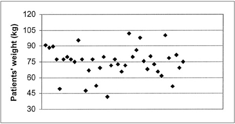
Fig. 1 Scatter graph depicting distribution of patients' weight.
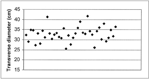
Fig. 2 Scatter graph depicting distribution of patients' transverse diameter.
Reference
-
1. Brenner DJ, Elliston CD, Hall EJ, Berdon W. Estimated risks of radiation-induced fatal cancer from pediatric CT. AJR Am J Roentgenol. 2001. 176:289–296.2. Nickoloff EL, Alderson PO. Radiation exposure to patients from CT: reality, public perception, and policy. AJR Am J Roentgenol. 2001. 176:285–287.3. United Nations Scientific Committee on the Effects of Atomic Radiation. UNSCEAR 1993 report. Sources and effects of ionizing radiation. United Nations, New York:4. Rehani MM, Berry M. Radiation doses in computed tomography. The increasing doses of radiation need to be controlled. BMJ. 2000. 320:593–594.5. Rehani MM, Bongartz G, Kalender W, et al. Managing X-ray dose in Computed Tomography. ICRP Special Task Force Report. 2000.6. McCollough CH, Zink FE. Performance evaluation of a multislice CT system. Med Phys. 1996. 26:2223–2230.7. Toth TL, Bromberg NB, Pan TS, et al. A dose reduction x-ray beam positioning system for high-speed multislice CT scanners. Med Phys. 2000. 27:2659–2668.8. Kalender WA, Wolf H, Suess C. Dose reduction in CT by anatomically adapted tube current modulation. II. Phantom measurements. Med Phys. 1999. 26:2248–2253.9. Kalra MK, Wittram C, Maher MM, et al. Can noise reduction filters improve low radiation dose chest CT images? Pilot Study. Radiology. 2003. 228:257–264.10. Kalra MK, Maher MM, Sahani DV, et al. Low-dose abdominal CT: Evaluation of image improvement from noise reduction filters-pilot study. Radiology. 2003. 228:251–256.11. Cohnen M, Fischer H, Hamacher J, Lins E, Kotter R, Modder U. CT of the head by use of reduced current and kilovoltage: relationship between image quality and dose reduction. AJNR Am J Neuroradiol. 2000. 21:1654–1660.12. Haaga JR, Miraldi F, MacIntyre W, LiPuma JP, Bryan PJ, Wiesen E. The effect of mAs variation upon computed tomography image quality as evaluated by in vivo and in vitro studies. Radiology. 1981. 138:449–454.13. Haaga JR. Radiation Dose Management: weighing risk versus benefit. AJR Am J Roentgenol. 2001. 177:289–291.14. Vade A, Demos TC, Olson MC, et al. Evaluation of image quality using 1:1 pitch and 1.5:1 pitch helical CT in children: a comparative study. Pediatr Radiol. 1996. 26:891–893.15. Diel J, Perlmutter S, Venkataramanan N, Mueller R, Lane MJ, Katz DS. Unenhanced helical CT using increased pitch for suspected renal colic: an effective technique for radiation dose reduction? J Comput Assist Tomogr. 2000. 24:795–801.16. Naidich DP, Marshall CH, Gribbin C, Arams RS, McCauley DI. Low-dose CT of the lungs: preliminary observations. Radiology. 1990. 175:729–731.17. Donnelly LF, Emery KH, Brody AS, et al. Minimizing radiation dose for pediatric body applications of single-detector helical CT: Strategies at a Large Children's Hospital. AJR Am J Roentgenol. 2001. 176:303–306.18. Han TS, van Leer EM, Seidell JC, Lean ME. Waist circumference action levels in the identification of cardiovascular risk factors: prevalence study in a random sample. Br Med J. 1995. 311:1401–1405.19. Seidell JC, Oosterlee A, Deurenberg P, Hautvast JG, Ruijs JH. Abdominal fat depots measured with computed tomography. Eur J Clin Nutr. 1998. 42:805–815.
- Full Text Links
-
- Actions
-
Cited
- CITED
-
- Close
- Share
-
- Similar articles
-
- The Optimal Parameter for Radiation Dose in Pediatric Low Dose Abdominal CT: Cross-sectional Dimensions versus Body Weight
- Radiation Dose and Imaging Quality of Abdominal Computed Tomography before and after Scan Protocol Adjustment: Single-Institution Experience in Three Years
- Comparison of Radiation Dose and Image Quality between the 2nd Generation and 3rd Generation Dual-Source Single-Energy and Dual-Source Dual-Energy CT of the Abdomen
- Performance of Half-dose Chest Computed Tomography in Lung Malignancy Using an Iterative Reconstruction Technique
- Evaluation of the effective dose and image quality of low-dose multi-detector CT for orthodontic treatment planning
