Cerebral Ischemia Detected with Diffusion-Weighted MR Imaging after Protected Carotid Artery Stenting: Comparison of Distal Balloon and Filter Device
- Affiliations
-
- 1Department of Radiology and Center for Imaging Science, Samsung Medical Center, Sungkyunkwan University School of Medicine, Seoul, Korea. hsbyun@smc.samsung.co.kr
- 2Stroke and Neurovascular Center, Samsung Medical Center, Sungkyunkwan University School of Medicine, Seoul, Korea.
- 3Department of Radiology, Konkuk University Hospital, Seoul, Korea.
- 4Department of Neurology, Samsung Medical Center, Sungkyunkwan University School of Medicine, Seoul, Korea.
- 5Division of Vascular Surgery, Department of Surgery, Samsung Medical Center, Sungkyunkwan University School of Medicine, Seoul, Korea.
- KMID: 1110724
- DOI: http://doi.org/10.3348/kjr.2007.8.4.276
Abstract
OBJECTIVE
The aim of this study was to examine the incidence of ischemia during protected carotid artery stenting (CAS) as well as to compare the protective efficacy of the balloon and filter devices on diffusion-weighted MR imaging (DWI). MATERIALS AND METHODS: Seventy-one consecutive protected CAS procedures in 70 patients with a severe (> 70%) or symptomatic moderate (> 50%) carotid artery stenosis were examined. A balloon device (PercuSurge GuardWire) and a filter device (FilterWire EX/EZ, Emboshield) was used in 33 cases (CAS-B group) and 38 cases (CAS-F group) to prevent distal embolization, respectively. All the patients underwent DWI within seven days before and after the procedures. The number of new cerebral ischemic lesions on the post-procedural DWI were counted and divided into ipsilateral and contralateral lesions according to the relationship with the stenting side. RESULTS: New cerebral ischemic lesions were detected in 13 (39.4%) out of the 33 CAS-Bs and in 15 (39.5%) out of the 38 CAS-Fs. The mean number of total, ipsilateral and contralateral new cerebral ischemic lesion was 2.39, 1.67 and 0.73 in the CAS-B group and 2.11, 1.32 and 0.79 in the CAS-F group, respectively. No statistical differences were found between the two groups (p = 0.96, 0.74 and 0.65, respectively). The embolic complications encountered included two retinal infarctions and one hemiparesis in the CAS-B group (9.09%), and one retinal infarction, one hemiparesis and one ataxia in the CAS-F group (7.89%). There was a similar incidence of embolic complications in the two groups (p = 1.00). CONCLUSION: The type of distal protection device used such as a balloon and filter does not affect the incidence of cerebral embolization after protected CAS.
Keyword
MeSH Terms
-
Adult
Aged
Aged, 80 and over
*Balloon Occlusion
Blood Vessel Prosthesis Implantation/*instrumentation
Brain Ischemia/*pathology
Carotid Stenosis/*surgery
*Diffusion Magnetic Resonance Imaging
Endarterectomy, Carotid/adverse effects/methods
Female
Humans
Intracranial Embolism/prevention & control
Male
Middle Aged
Paresis/etiology
Retinal Artery Occlusion/etiology
Severity of Illness Index
*Stents
Figure
-
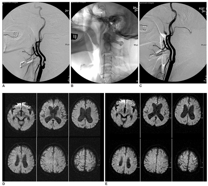
Fig. 1 A 72-year-old man who underwent protected carotid artery stenting with a balloon device. A. Pre-stenting angiogram shows severe stenosis (86.3%) at the left internal carotid artery. B. A balloon device is deployed in the distal carotid artery (arrow). C. After carotid artery stenting, the lumen of the left internal carotid artery is successfully dilated. D. No ischemic lesion is shown in bilateral cerebral hemispheres on the pre-stenting diffusion weighted MR imaging. E. Multiple small new hyperintensities are shown on the post-stenting diffusion-weighted MR imaging. Note the new hyperintnesities are distributed in not only the ipsilateral but also the contralateral cerebral hemisphere. However, no symptomatic neurological complications occurred after carotid artery stenting.
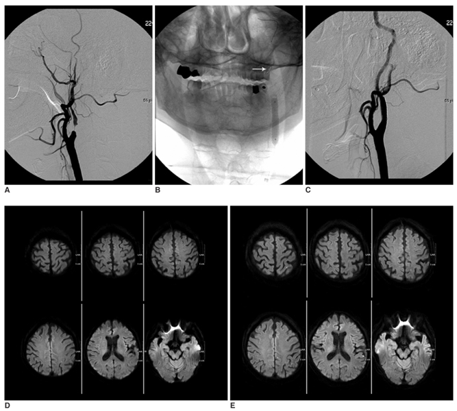
Fig. 2 A 71-year-old man who underwent protected carotid artery stenting with a filter device. A. Pre-stenting angiogram shows a severe string like stenosis of the left internal carotid artery. The post-stenotic distal internal carotid artery is also narrow compared with the external carotid artery (pseudo-occlusion). B. A filter device is deployed in the distal carotid artery (arrow). C. After carotid artery stenting, the lumen of the left internal carotid artery is successfully dilated. D. No ischemic lesion is shown in bilateral cerebral hemispheres on the pre-stenting diffusion weighted MR imaging. E. Multiple small new hyperintensities are observed in the ipsilateral cerebral hemisphere on the post-stenting diffusion-weighted MR images. However, no symptomatic neurological complications occurred after carotid artery stenting.
Cited by 2 articles
-
Relationship Between Nocturnal Dip, Carotid Artery Blood Flow, Brain Ischemic Change in Open Angle Glaucoma
Hong Ryung Seo, Sang Wook Jin, Sae Heun Rho
J Korean Ophthalmol Soc. 2013;54(9):1386-1394. doi: 10.3341/jkos.2013.54.9.1386.Ophthalmic Artery Occlusion After Carotid Revascularization
Yeon Jin Yi, Ji Kwang Yun, Dae Won Kim, Sung Don Kang
J Cerebrovasc Endovasc Neurosurg. 2013;15(4):326-329. doi: 10.7461/jcen.2013.15.4.326.
Reference
-
1. Lövblad KO, Laubach HJ, Baird AE, Curtin F, Schlaug G, Edelman RR, et al. Clinical experience with diffusion-weighted MR in patients with acute stroke. AJNR Am J Neuroradiol. 1998. 19:1061–1066.2. Beauchamp NJ Jr, Barker PB, Wang PY, vanZijl PC. Imaging of acute cerebral ischemia. Radiology. 1999. 212:307–324.3. van Everdingen KJ, van der Grond J, Kappelle LJ, Ramos LM, Mali WP. Diffusion-weighted magnetic resonance imaging in acute stroke. Stroke. 1998. 29:1783–1790.4. Forbes KP, Shill HA, Britt PM, Zabramski JM, Spetzler RF, Heiserman JE. Assessment of silent embolism from carotid endarterectomy by use of diffusion-weighted imaging: work in progress. AJNR Am J Neuroradiol. 2001. 22:650–653.5. Barth A, Remonda L, Lövblad KO, Schroth G, Seiler RW. Silent cerebral ischemia detected by diffusion-weighted MRI after carotid endarterectomy. Stroke. 2000. 31:1824–1828.6. Feiwell RJ, Besmertis L, Sarkar R, Saloner DA, Rapp JH. Detection of clinically silent infarcts after carotid endarterectomy by use of diffusion-weighted imaging. AJNR Am J Neuroradiol. 2001. 22:646–649.7. Rordorf G, Bellon RJ, Budzik RE Jr, Farkas J, Reinking GF, Pergolizzi RS, et al. Silent thromboembolic events associated with the treatment of unruptured cerebral aneurysms by use of Guglielmi detachable coils: prospective study applying diffusion-weighted imaging. AJNR Am J Neuroradiol. 2001. 22:5–10.8. Muller M, Reiche W, Langenscheidt P, Hassfeld J, Hagen T. Ischemia after carotid endarterectomy: comparison between transcranial Doppler sonography and diffusion-weighted MR imaging. AJNR Am J Neuroradiol. 2000. 21:47–54.9. Jaeger HJ, Mathias KD, Drescher R, Hauth E, Bockisch G, Demirel E, et al. Diffusion-weighted MR imaging after angioplasty or angioplasty plus stenting of arteries supplying the brain. AJNR Am J Neuroradiol. 2001. 22:1251–1259.10. Britt PM, Heiserman JE, Snider RM, Shill HA, Bird CR, Wallace RC. Incidence of postangiographic abnormalities revealed by diffusion-weighted MR imaging. AJNR Am J Neuroradiol. 2000. 21:55–59.11. Bendszus M, Koltzenburg M, Burger R, Warmuth-Metz M, Hofmann E, Solymosi L. Silent embolism in diagnostic cerebral angiography and neurointerventional procedures: a prospective study. Lancet. 1999. 354:1594–1597.12. Hammer FD, Lacroix V, Duprez T, Grandin C, Verhelst R, Peeters A, et al. Cerebral microembolization after protected carotid artery stenting in surgical high-risk patients: results of a 2-year prospective study. J Vasc Surg. 2005. 42:847–853. discussion 853.13. du Mesnil de Rochemont R, Schneider S, Yan B, Lehr A, Sitzer M, Berkefeld J. Diffusion-weighted MR imaging lesions after filter-protected stenting of high-grade symptomatic carotid artery stenoses. AJNR Am J Neuroradiol. 2006. 27:1321–1325.14. Asakura F, Kawaguchi K, Sakaida H, Toma N, Matsushima S, Kuraishi K, et al. Diffusion-weighted magnetic resonance imaging in carotid angioplasty and stenting with balloon embolic protection devices. Neuroradiology. 2006. 48:100–112.15. Asakura F, Kawaguchi K, Sakaida H, Toma N, Matsushima S, Kuraishi K, et al. Diffusion-weighted MR imaging in carotid angioplasty and stenting with protection by the reversed carotid arterial flow. AJNR Am J Neuroradiol. 2006. 27:753–758.16. Gröschel K, Ernemann U, Riecker A, Schmidt F, Terborg C, Kastrup A. Incidence and risk factors for medical complications after carotid artery stenting. J Vasc Surg. 2005. 42:1101–1106. discussion 1106-1107.17. Henry M, Gopalakrishnan L, Rajagopal S, Rath PC, Henry I, Hugel M. Bilateral carotid angioplasty and stenting. Catheter Cardiovasc Interv. 2005. 64:275–282.18. Hobson RW, Howard VJ, Roubin GS, Brott TG, Ferguson RD, Popma JJ, et al. Carotid artery stenting is associated with increased complications in octogenarians: 30-day stroke and death rates in the CREST lead-in phase. J Vasc Surg. 2004. 40:1106–1111.19. Kastrup A, Gröschel K, Krapf H, Brehm BR, Dichgans J, Schulz JB. Early outcome of carotid angioplasty and stenting with and without cerebral protection devices: a systematic review of the literature. Stroke. 2003. 34:813–819.20. Kastrup A, Gröschel K, Schulz JB, Nägele T, Ernemann U. Clinical predictors of transient ischemic attack, stroke, or death within 30 days of carotid angioplasty and stenting. Stroke. 2005. 36:787–791.21. Krapf H, Nägele T, Kastrup A, Bühring U, Grönewäller E, Skalej M, et al. Risk factors for periprocedural complications in carotid artery stenting without filter protection: a serial diffusion-weighted MRI study. J Neurol. 2006. 253:364–371.22. Lanzer P, Weser R, Prettin C. Carotid-artery stenting in a high-risk patient population-single centre, single operator results. Clin Res Cardiol. 2006. 95:4–12.23. Reiter M, Bucek RA, Effenberger I, Boltuch J, Lang W, Ahmadi R, et al. Plaque echolucency is not associated with the risk of stroke in carotid stenting. Stroke. 2006. 37:2378–2380.24. Roubin GS, New G, Iyer SS, Vitek JJ, Al-Mubarak N, Liu MW, et al. Immediate and late clinical outcomes of carotid artery stenting in patients with symptomatic and asymptomatic carotid artery stenosis: a 5-year prospective analysis. Circulation. 2001. 103:532–537.25. Spagnoli LG, Mauriello A, Sangiorgi G, Fratoni S, Bonanno E, Schwartz RS, et al. Extracranial thrombotically active carotid plaque as a risk factor for ischemic stroke. JAMA. 2004. 292:1845–1852.26. Sztriha LK, Vörös E, Sas K, Szentgyörgyi R, Pócsik A, Barzó P, et al. Favorable early outcome of carotid artery stenting without protection devices. Stroke. 2004. 35:2862–2866.27. Theiss W, Hermanek P, Mathias K, Ahmadi R, Heuser L, Hoffmann FJ, et al. Pro-CAS: a prospective registry of carotid angioplasty and stenting. Stroke. 2004. 35:2134–2139.28. Timaran CH. Clinical predictors of transient ischemic attack, stroke, or death within 30 days of carotid angioplasty and stenting. Perspect Vasc Surg Endovasc Ther. 2005. 17:384–385.29. Wholey MH, Al-Mubarek N, Wholey MH. Updated review of the global carotid artery stent registry. Catheter Cardiovasc Interv. 2003. 60:259–266.30. Hofmann R, Niessner A, Kypta A, Steinwender C, Kammler J, Kerschner K, et al. Risk score for peri-interventional complications of carotid artery stenting. Stroke. 2006. 37:2557–2561.31. Endovascular versus surgical treatment in patients with carotid stenosis in the Carotid and Vertebral Artery Transluminal Angioplasty Study (CAVATAS): a randomised trial. Lancet. 2001. 357:1729–1737.32. Roh HG, Byun HS, Ryoo JW, Na DG, Moon WJ, Lee BB, et al. Prospective analysis of cerebral infarction after carotid endarterectomy and carotid artery stent placement by using diffusion-weighted imaging. AJNR Am J Neuroradiol. 2005. 26:376–384.33. Yadav JS, Wholey MH, Kuntz RE, Fayad P, Katzen BT, Mishkel GJ, et al. Protected carotid-artery stenting versus endarterectomy in high-risk patients. N Engl J Med. 2004. 351:1493–1501.34. Ouriel K, Wholey MH, Fayad P, Katzen BT, Whitlow P, Frentzko M, et al. Feasibility trial of carotid stenting with and without an embolus protection device. J Endovasc Ther. 2005. 12:525–537.35. Carlino M, De Gregorio J, Di Mario C, Anzuini A, Airoldi F, Albiero R, et al. Prevention of distal embolization during saphenous vein graft lesion angioplasty. Experience with a new temporary occlusion and aspiration system. Circulation. 1999. 99:3221–3223.36. Theron J, Courtheoux P, Alachkar F, Bouvard G, Maiza D. New triple coaxial catheter system for carotid angioplasty with cerebral protection. AJNR Am J Neuroradiol. 1990. 11:869–874.37. Henry M, Polydorou A, Henry I, Polydorou I, Hugel IM, Anagnostopoulou S. Angioplasty and stenting of extracranial vertebral artery stenosis. Int Angiol. 2005. 24:311–324.38. Bogousslavsky J, Regli F, Hungerbuhler JP, Chrzanowski R. Transient ischemic attacks and external carotid artery. A retrospective study of 23 patients with an occlusion of the internal carotid artery. Stroke. 1981. 12:627–630.39. Mames RN, Snady-McCoy L, Guy J. Central retinal and posterior ciliary artery occlusion after particle embolization of the external carotid artery system. Ophthalmology. 1991. 98:527–531.40. Ohki T, Parodi J, Veith FJ, Bates M, Bade M, Chang D, et al. Efficacy of a proximal occlusion catheter with reversal of flow in the prevention of embolic events during carotid artery stenting: an experimental analysis. J Vasc Surg. 2001. 33:504–509.41. Zahn R, Ischinger T, Mark B, Gass S, Zeymer U, Schmalz W, et al. Embolic protection devices for carotid artery stenting: is there a difference between filter and distal occlusive devices? J Am Coll Cardiol. 2005. 45:1769–1774.42. Müller-Hülsbeck S, Jahnke T, Liess C, Glass C, Paulsen F, Grimm J, et al. In vitro comparison of four cerebral protection filters for preventing human plaque embolization during carotid interventions. J Endovasc Ther. 2002. 9:793–802.43. Al-Mubarak N, Colombo A, Gaines PA, Iyer SS, Corvaja N, Cleveland TJ, et al. Multicenter evaluation of carotid artery stenting with a filter protection system. J Am Coll Cardiol. 2002. 39:841–846.44. Angelini A, Reimers B, Della Barbera M, Sacca S, Pasquetto G, Cernetti C, et al. Cerebral protection during carotid artery stenting: collection and histopathologic analysis of embolized debris. Stroke. 2002. 33:456–461.45. Whitlow PL, Lylyk P, Londero H, Mendiz OA, Mathias K, Jaeger H, et al. Carotid artery stenting protected with an emboli containment system. Stroke. 2002. 33:1308–1314.46. Cremonesi A, Manetti R, Setacci F, Setacci C, Castriota F. Protected carotid stenting: clinical advantages and complications of embolic protection devices in 442 consecutive patients. Stroke. 2003. 34:1936–1941.47. Castellan L, Causin F, Danieli D, Perini S. Carotid stenting with filter protection. Correlation of ACT values with angiographic and histopathologic findings. J Neuroradiol. 2003. 30:103–108.48. White CJ, Iyer SS, Hopkins LN, Katzen BT, Russell ME. Carotid stenting with distal protection in high surgical risk patients: the BEACH trial 30 day results. Catheter Cardiovasc Interv. 2006. 67:503–512.49. Hill MD, Morrish W, Soulez G, Nevelsteen A, Maleux G, Rogers C, et al. Multicenter evaluation of a self-expanding carotid stent system with distal protection in the treatment of carotid stenosis. AJNR Am J Neuroradiol. 2006. 27:759–765.
- Full Text Links
-
- Actions
-
Cited
- CITED
-
- Close
- Share
-
- Similar articles
-
- Massive Cerebral Microemboli after Protected Carotid Artery Angioplasty and Stenting Using a Distal Filter Embolic Protection Device for a Vulnerable Plaque with a Lipid Rich Necrotic Core and Intraplaque Hemorrhage: A Case Report
- Clinical Analysis Comparing Efficacy between a Distal Filter Protection Device and Proximal Balloon Occlusion Device during Carotid Artery Stenting
- Central Retinal Artery Occlusion After Carotid Artery Angioplasty and Stenting in an Elderly Patient: A Case Report
- Diffusion-Weighted MR Imaging after Carotid Artery Stenting
- Early Detection of Hyperacute Cerebral Infarction in Dogs: Comparison of Unenhanced CT, Diffusion-weighted,Spin-echo T2 - weighted, and Fast FLAIR MR Imaging
