Catheter-guided percutaneous heartworm removal using a nitinol basket in dogs with caval syndrome
- Affiliations
-
- 1Section of Small Animal Internal Medicine, and Institute of Veterinary Science, College of Veterinary Medicine, Kangwon National University, Chuncheon 200-701, Korea. hyun5188@kangwon.ac.kr
- KMID: 1106565
- DOI: http://doi.org/10.4142/jvs.2011.12.2.199
Abstract
- Carval syndrome is a severe heartworm infection where the worms have migrated to the right atrium and vena cava; this condition is associated with a myriad of clinical signs. Several non-surgical and interventional methods are currently used for mechanical worm removal. However, the success rate and complications related to these methods are heavily dependent on methodology and retrieval devices used. In this study, we developed a catheter-guided heartworm removal method using a retrieval basket that can easily access pulmonary arteries and increase the number of worms removed per procedure. With this technique, we successfully treated four dogs with caval syndrome.
Keyword
MeSH Terms
Figure
-

Fig. 1 Heartworm removal procedure. After induce surgical anesthesia, the left jugular vein was exposed. An introducer sheath was inserted into the left external jugular vein guided by the pre-placed guide wire into the right atrium, right ventricle, and pulmonary artery (A). The basket device was inserted into the introducer and spread-folded to catch heartworms under fluoroscopic guidance (B and C).
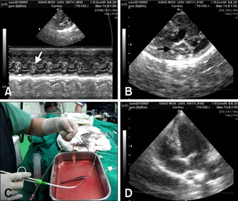
Fig. 2 Echocardiography and clinical outcomes. (A) M-mode echocardiography revealed clumps (arrow) of heartworms in the right ventricle due to heavy worm burden. (B) B-mode echocardiography showed heartworms migrating from the right ventricle to the right atrium (arrow). (C) Multiple worm removal per trial in a dog (Case 2). (D) Disappearance of heartworms from the right atrium and ventricle after the removal procedure on B-mode echocardiography in a dog (Case 1).
Reference
-
1. Arita N, Yamane I, Takemura N. Comparison of canine heartworm removal rates using flexible alligator forceps guided by transesophageal echocardiography and fluoroscopy. J Vet Med Sci. 2003. 65:259–261.
Article2. Atkins C. Ettinger SJ, Feldman EC, editors. Canine heartworm disease. Textbook of Veterinary Internal Medicine. 2005. 6th ed. St. Louis: Saunders;1118–1136.3. Kittleson MD. Kittleson MD, Kienle RD, editors. Heartworm infestation and disease (dirofilariasis). Small Animal Cardiovascular Medicine. 1998. 1st ed. St. Louis: Mosby;370–401.4. Salimi N, Mahajan A, Don J, Schwartz B. A novel stone retrieval basket for more efficient lithotripsy procedures. J Med Eng Technol. 2009. 33:142–150.
Article
- Full Text Links
-
- Actions
-
Cited
- CITED
-
- Close
- Share
-
- Similar articles
-
- Percutaneous heartworm removal from dogs with severe heart worm (Dirofilaria immitis) infestation
- Evaluation of improved transvenous heartworm extraction brush in dogs with caval syndrome
- Modeling of transmission pathways on canine heartworm dynamics
- Percutaneous transhepatic removal of common bile duct stone: a case report
- A survey of canine heartworm infections among German shepherds in South Korea
