J Vet Sci.
2008 Jun;9(2):193-196. 10.4142/jvs.2008.9.2.193.
Contribution of the xenograft bone plate-screw system in lumbar transpedicular stabilization of dogs: an in-vitro study
- Affiliations
-
- 1Department of Surgery, Faculty of Veterinary Medicine, Uludag University, Bursa, Turkey. hsalci@uludag.edu.tr
- 2Department of Neurosurgery, Faculty of Medicine, Uludag University, Bursa, Turkey.
- 3Department of Machines, Faculty of Engineering and Architecture, Uludag University, Bursa, Turkey.
- KMID: 1106235
- DOI: http://doi.org/10.4142/jvs.2008.9.2.193
Abstract
- We performed biomechanical comparison of a xenograft bone plate-screw (XBPS) system for achieving cadaveric lumbar transpedicular stabilization (TS) in dogs. Twenty dogs' cadaveric L2-4 lumbar specimens were harvested and their muscles were removed, but the discs and ligaments were left intact. These specimens were separated to four groups: the L2-4 intact group as control (group I, n = 5), the L3 laminectomy and bilateral facetectomy group (LBF) (group II, n = 5), the LBF plus TS with metal plate-screw group (group III, n = 5) and the LBF plus TS with XBPS group (group IV, n = 5). Five kinds of biomechanical tests were applied to the specimens: flexion, extension, left-right bending and rotation. The averages of the 16 stiffness values were calculated and then these were statistically analyzed. The statistical results show that the XBPS system contributes spinal stability and this system can be a good choice for achieving TS.
MeSH Terms
Figure
-
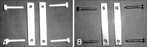
Fig. 1 (A) Xenograft bone plates and screws used for transpedicular stabilization. (B) Metal plates and screws used for transpedicular stabilization.
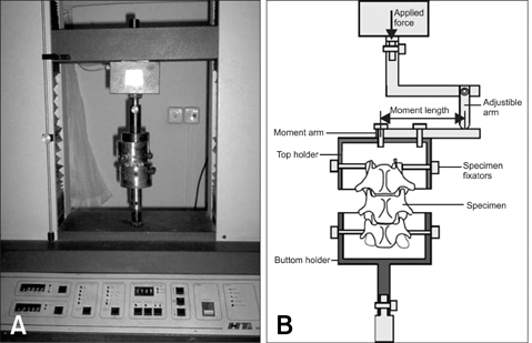
Fig. 2 (A) View of a tensile-compression testing machine used for the biomechanical tests. (B) Schematic of the experiment.
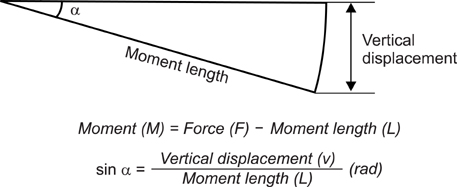
Fig. 3 Calculation of radian angles for converting the forces to the moments.
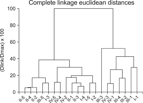
Fig. 4 The results of the Euclidian distance method and the four main clusters.
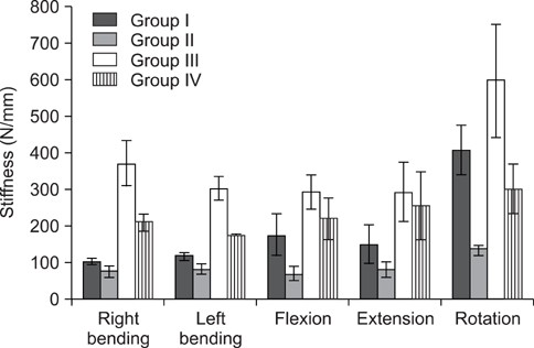
Fig. 5 The comparison of stiffness for xenograft bone platescrew following biomechanical test.
Reference
-
1. Benzel EC. Biomechanics of Spine Stabilization: Principles and Clinical Practice. 1995. New York: McGraw-Hill;89–107.2. Boucher HH. A method of spinal fusion. J Bone Joint Surg. 1959. 41B:248–249.
Article3. Brodke DS, Bachus KN, Mohr RA, Nguyen BK. Segmental pedicle screw fixation or cross-links in multilevel lumbar constructs. a biomechanical analysis. Spine J. 2001. 1:373–379.
Article4. Harrington PR. The management of scoliosis by spine instrumentation: An evaluation of more than 200 cases. South Med J. 1963. 56:1367–1377.5. Hasegawa K, Takahashi HE, Uchiyama S, Hirano T, Hara T, Washio T, Sugiura T, Youkaichiya M, Ikeda M. An experimental study of a combination method using a pedicle screw and laminar hook for the osteoporotic spine. Spine. 1997. 22:958–962.
Article6. Johnsson R, Axelsson P, Gunnarsson G, Strömqvist B. Stability of lumbar fusion with transpedicular fixation determined by roentgen stereophotogrammetric analysis. Spine. 1994. 24:687–690.
Article7. Kumano K, Hirabayashi S, Ogawa Y, Aota Y. Pedicle screws and bone mineral density. Spine. 1994. 19:1157–1161.
Article8. Myers BS, Belmont PJ Jr, Richardson WJ, Yu JR, Harper KD, Nightingale RW. The role of imaging and In Situ biomechanical testing in assessing pedicle screw pull-out strength. Spine. 1996. 21:1962–1968.
Article9. Sandén B, Olerud C, Petrén-Mallmin M, Larsson S. Hydroxyapatite coating improves fixation of pedicle screws. A clinical study. J Bone Joint Surg Br. 2002. 84:387–391.10. White AA, Panjabi MM. Clinical Biomechanics of the Spine. 1990. Philadelphia: Lippincott;84–126.
- Full Text Links
-
- Actions
-
Cited
- CITED
-
- Close
- Share
-
- Similar articles
-
- Loss of the Sagittal angle in the Instrumented Segments after Pedicular Screw Fixation of the Degenerative Lumbar Diseases
- Posterior Lower Cervical fixation with the AME Haid Universal Bone Plate System
- Follow-up Result of Transpedicular Screw Fixation in Lumbar Spinal Stenosis and Spondylolisthesis
- Operative Treatment for Hangman Fracture of the Axis: Review of Fixation Methods and Indications
- Instrument-Related Complications Following Lumbar Transpedicular Screw Fixation
