Korean J Ophthalmol.
2007 Mar;21(1):61-64. 10.3341/kjo.2007.21.1.61.
Acute Angle-Closure Glaucoma from Spontaneous Massive Hemorrhagic Retinal Detachment
- Affiliations
-
- 1Department of Ophthalmology, Hanyang University, School of Medicine, Hanyang University Guri Hospital, Gyeonggi-do, Korea. lyjot@hanyang.ac.kr
- KMID: 1105034
- DOI: http://doi.org/10.3341/kjo.2007.21.1.61
Abstract
- PURPOSE: To report a case of acute angle-closure glaucoma resulting from spontaneous hemorrhagic retinal detachment. METHODS: An 81-year-old woman visited our emergency room for severe ocular pain and vision loss in her left eye. Her intraocular pressures (IOPs) were 14 mmHg in the right eye and 58 mmHg in the left eye. Her visual acuity was 0.4 in the right eye but she had no light perception in the left eye. The left anterior chamber depth was shallow and gonioscopy of the left eye showed a closed angle. In comparison, the right anterior chamber depth was normal and showed a wide, open angle. Computed tomography and ultrasonography demonstrated retinal detachment due to subretinal hemorrhage. After systemic and topical antiglaucoma medications failed to relieve her intractable severe ocular pain, she underwent enucleation. RESULTS: The ocular pathology specimen showed that a large subretinal hemorrhage caused retinal detachment and pushed displaced the lens-iris diaphragm, resulting in secondary angle-closure glaucoma. CONCLUSIONS: Prolonged anticoagulant therapy may cause hemorrhagic retinal detachment and secondary angle-closure glaucoma. If medical therapy fails to relieve pain or if there is suspicion of an intraocular tumor, enucleation should be considered as a therapeutic option.
MeSH Terms
Figure
-
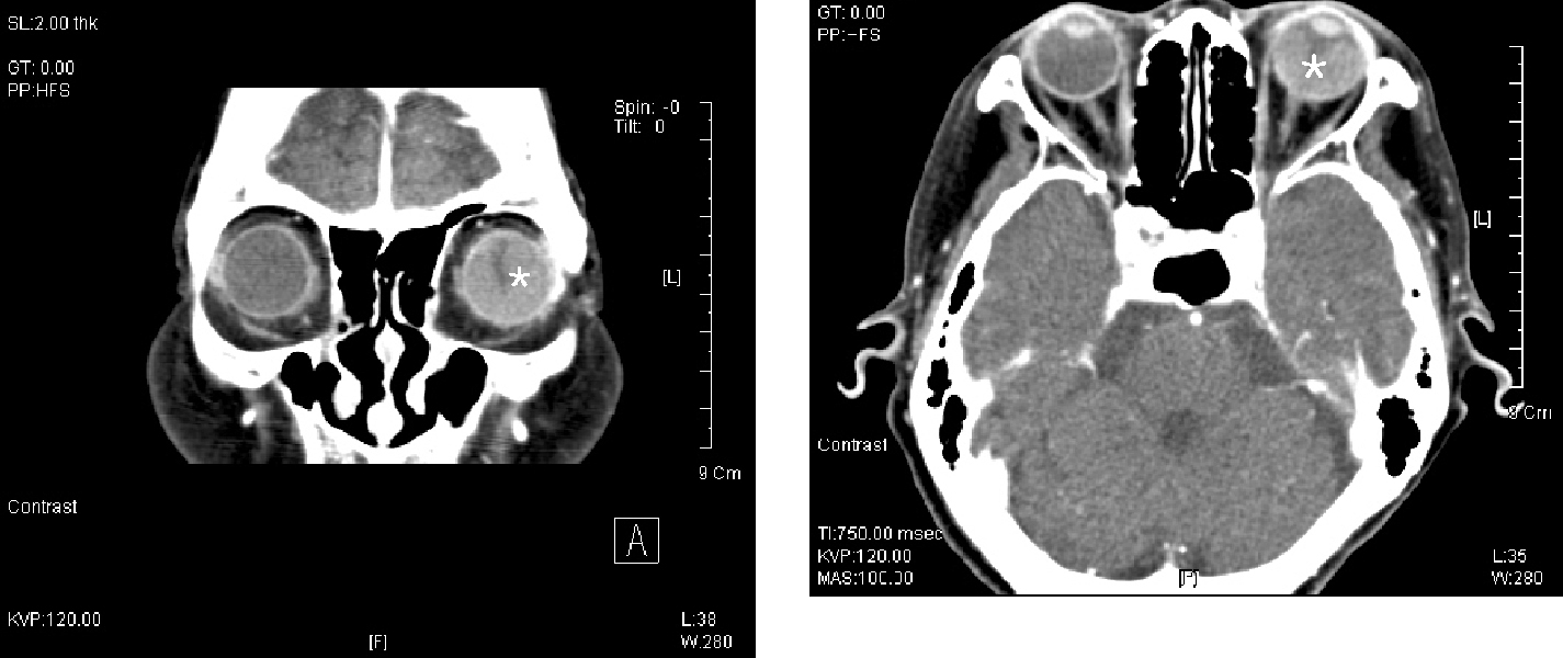
Fig. 1 Orbital computed tomography with contrast enhancement showed a large hemorrhagic retinal detachment (asterisk) in the left eye.
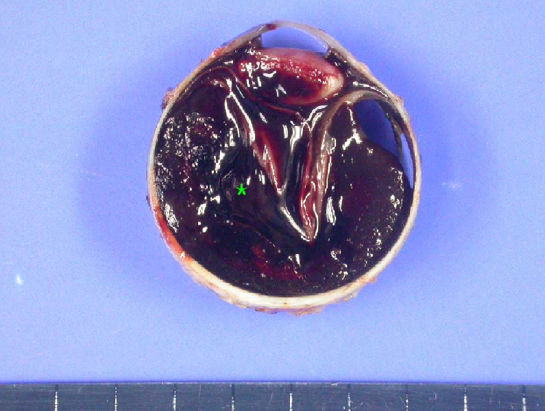
Fig. 2 The firm left globe was horizontally sectioned. Massive amounts of degenerated blood (asterisk) filled the subretinal space. The totally detached retina touched the posterior surface of the lens and the anterior chamber was shallow.
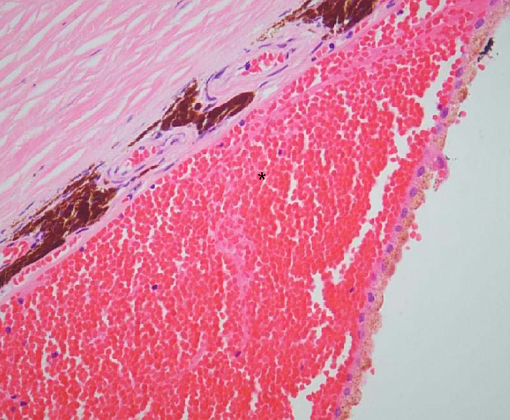
Fig. 3 Hemorrhagic RPE detachment was found. Blood also filled the subretinal space (asterisk) (hematoxylin-eosin stain: original magnification, ×400).
Reference
-
1. Brown GC, Tasman WS, Shields JA. Massive subretinal hemorrhage and anticoagulant therapy. Can J Ophthalmol. 1982. 17:227–230.2. Mosley DH, Schatix IJ, Breneman GM, Keyes JW. Long-term anticoagulant therapy. JAMA. 1963. 186:914–916.3. Pollard JW, Hamilton MJ, Christensen NA, Achor R WP. Problems associated with long-term anticoagulant therapy. Circulation. 1962. 25:311–317.4. Edward P. Massive choroidal hemorrhage in age-related macular degeneration : a complication of anticoagulant therapy. J Am Optom Assoc. 1997. 67:223–226.5. Gordan DM, Mead G. Retinal hemorrhage with visual loss during anticoagulant therapy: case report. J Am Geriatr Soc. 1968. 16:99–100.6. Pesin SR, Katz LJ, Augsburger JJ, et al. Acute angle-closure glaucoma from spontaneous massive hemorrhagic retinal or choroidal detachment. Ophthalmology. 1990. 97:76–84.7. Parson JH. The pathology of the eye. 1906. Vol. 1:1st ed. New York: GP Putnam's Sons;1087–1088.8. Chen SN, Ho CL, Ho JD, et al. Acute angle-closure glaucoma resulting from spontaneous hemorrhagic retinal detachment in age-related macular degeneration; case reports and literature review. Jpn J Ophthalmol. 2001. 45:270–275.9. Wood WJ, Smith TR. Senile disciform macular degeneration complicated by massive hemorrhagic retinal detachment and angle-closure glaucoma. Retina. 1983. 3:296–303.10. Feman SS, Bartlett RE, Roth AM, Foos RY. Intraocular hemorrhage and blindness associated with systemic anticoagulation. JAMA. 1972. 220:1354–1355.11. Steiemann T, Goins K, Smith T, et al. Acute angle closure glaucoma complicating hemorrhagic choroidal detachment associated with parenteral thrombolytic agents. Am J Ophthalmol. 1988. 106:752–753.12. Kozlowski IMD, Hirose T, Jalkh AE. Massive subretinal hemorrhage with acute angle-closure glaucoma in chronic myelocytic leukemia. Am J Ophthalmol. 1987. 103:832–833.13. Caronia RM, Sturm RT, Fastenberg DM, et al. Bilateral secondary angle-closure glaucoma as a complication in nonophthalmic patient. Am J Ophthalmol. 1998. 126:307–309.14. El Baba F, Jarrett WH II, Harbin TS, et al. Massive hemorrhage complicating age related macular degeneration. Ophthalmology. 1986. 93:1581–1592.15. Butner RW, McPherson AR. Spontaneous vitreous hemorrhage. Ann Ophthalmol. 1982. 14:268–270.16. Oyakawa RT, Michels RG, Blasé WP. Vitrectomy for nondiabetic vitreous hemorrhage. Am J Ophthalmol. 1983. 96:517–525.17. Pastror BH, Resnick ME, Rodman T. Serious hemorrhagic complications of anticoagulant therapy. JAMA. 1962. 25:311–317.18. Yanoff M. Glaucoma mechanisms in ocular malignant melanomas. Am J Ophthalmol. 1970. 70:898–904.19. Chu TG, Cano MR, Green RL, et al. Massive suprachoroidal hemorrhage with central retinal apposition. A clinical and echographic study. Arch Ophthalmol. 1991. 109:1575–1581.20. Knopp EA, Chymm KY. Spontaneous expulsive choroidal hemorrhage: CT findings. Am J Neuroradiol. 1990. 11:1208–1209.
- Full Text Links
-
- Actions
-
Cited
- CITED
-
- Close
- Share
-
- Similar articles
-
- A Case of Acute Angle-closure Glaucoma Secondary to Spontaneous Suprachoroidal Hemorrhage
- Angle-closure Attack after Retinal Pigment Epithelium Double-tear and Hemorrhagic Retinal Detachment in Exudative Macular Degeneration
- A Case of Bilateral Malignant Glaucoma with Ciliochoroidal Detachment
- Bilateral Acute Angle-Closure Glaucoma after Macular Hole Surgery
- Biometric Comparisons between Acute and Chronic Angle Closure Glaucoma
