Diprosopus, craniorachischisis, arthrogryposis, and other associated anomalies in a stillborn lamb
- Affiliations
-
- 1Department of Reproduction, Faculty of Veterinary Medicine, Kafkas University, Kars 36100, Turkey.
- 2Department of Pathology, Faculty of Veterinary Medicine, Kafkas University, Kars 36100, Turkey. kadirozcan36@hotmail.com
- 3Department of Anatomy, Faculty of Veterinary Medicine, Kafkas University, Kars 36100, Turkey.
- 4Department of Reproduction, Faculty of Veterinary Medicine, Erciyes University, Kayseri 38090, Turkey.
- KMID: 1104921
- DOI: http://doi.org/10.4142/jvs.2008.9.4.429
Abstract
- Congenital malformations with multiple anomalies have been described infrequently in the veterinary literature. A stillborn male crossbred lamb with diprosopus, craniorachischisis, and arthrogryposis was examined macroscopically and histopathologically in this study. The left head was smaller than the right head. Micrencephaly, agnathia, and a rudimentary tongue, which was adherent to the palate, were present in the left head. Micrencephaly, brachygnathia superior, and cleft palate were present in the right head. Cerebellar agenesis and spinal cord hypoplasia were observed. The cerebrums and the spinal cord were covered with a tapering membranous structure. Neural and dermal tissues were noted to intervene upon microscopic examination of this structure. Disorganization of neurons was observed in both cerebrums, though it was more severe in the left one. This case demonstrates many congenital defects occurring together in a lamb.
Keyword
MeSH Terms
Figure
-
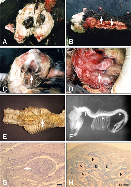
Fig. 1 (A) Frontal view of the diprosopic lamb. (B) Craniorachischisis. A tapering membranous structure covering the spinal cord (arrows). (C) Agnathia in the left head. Skin covering the presumptive location of the lower jaw (arrow). (D) A rudimentary tongue, which is adherent to the palate, in the left head (arrow). (E) Dorsal view of the lamb's vertebral column. Severe deformation in the vertebral column. Scoliosis in the thoracal vertebrae (arrow). (F) Lateral radiograph of the lamb. Kyphosis in the cervical, thoracal, and lumbar vertebrae (arrows). (G) Microscopic view of disorganization in the left brain. Fibrous tissue in the brain (arrow). Masson's trichrome. Scale bar = 56 µm. (H) Intervention of the neural (N) and dermal (D) tissues under the membranous structure. Masson's trichrome. Scale bar = 140 µm.
Reference
-
1. Canda MŞ, Canda T. Temel patoloji 4: Nöropatoloji. 1992. Bornova: Ege Üniversitesi Yayını;13–20.2. Dennis SM. Perinatal lamb mortality in western Australia. 7. Congenital defects. Aust Vet J. 1975. 51:80–82.
Article3. Dennis SM, Leipold HW. Ovine congenital defects. Vet Bull. 1979. 49:233–239.4. Dozsa L. A case of rare monstrosity in a calf. Pathol Vet. 1966. 3:226–233.
Article5. Fisher KRS, Partlow GD, Walker AF. Clinical and anatomical observations of a two-headed lamb. Anat Rec. 1986. 214:432–440.
Article6. Hartley WJ, Haughley KG. An outbreak of micrencephaly in lambs in New South Wales. Aust Vet J. 1974. 50:55–58.
Article7. Hiraga T, Dennis SM. Dennis SM, editor. Congenital duplication. The Veterinary Clinics of North America, Food Animal Practice. Congenital Abnormalities. 1993. Philadelphia: Saunders;145–161.
Article8. Leipold HW, Dennis SM. Dicephalus in two calves. Am J Vet Res. 1972. 33:421–423.9. Leipold HW, Dennis SM. Diprosopus in newborn calves. Cornell Vet. 1972. 62:282–288.10. Madarame H, Ito N, Takai S. Dicephalus, Arnold-chiari malformation and spina bifida in a Japanese black calf. J Vet Med A. 1993. 40:155–160.
Article11. Mazzulo G, Germanà A, De Vico G, Germanà G. Diprosopiasis in a lamb. A case report. Anat Histol Embryol. 2003. 32:60–62.
Article12. McGirr WJ, Partlow GD, Fisher KRS. Two-headed, two-necked conjoined twin calf with partial duplication of thoracoabdominal structures: Role of blastocyst hatching. Anat Rec. 1987. 217:196–202.
Article13. Moerman P, Fryns JP, Goddeeris P, Lauweryns JM, Van Assche A. Aberrant twinning (Diprosopus) associated with anencephaly. Clin Genet. 1983. 24:252–256.
Article14. Noden DM, De Lahunta A. The Embryology of Domestic Animals: Developmental Mechanisms and Malformations. 1985. Baltimore: Williams & Wilkins;109–152.15. Ozcan K, Ozturkler Y, Sozmen M, Takci I. Diprosopus in a cross bred calf. Indian Vet J. 2005. 82:650–651.16. Roberts SJ. Veterinary Obstetrics and Genital Disease (Theriogenelogy). 1986. 3th ed. Woodstock: Edwards Brothers;51–91.17. Saperstein G. Diprosopus in a hereford calf. Vet Rec. 1981. 108:234–235.
Article18. Sönmez G, Özbilgin S, Serbest A, Mısırlıoğlu D. A case of diprosopus in a kid. Uludağ Üniv Vet Fak Derg. 1992. 11:93–98.19. Türkütanıt SS, Sağlam YS, Bozoğlu H. Diprosopus in a calf. İstanbul Üniv Vet Fak Derg. 1996. 22:253–256.
