J Vet Sci.
2006 Mar;7(1):83-85. 10.4142/jvs.2006.7.1.83.
Histology of two rice bodies isolated from the stifle of an adult draught horse stallion
- Affiliations
-
- 1Institute of General Anaesthesiology and Surgical Pathology of Large Animals, University of Liege, Bat B 6a (CORD/ Chimie), 4000 Sart Tilman, Belgium. ni.schneider@gmx.net
- 2Center of Oxygen, Research and Development, University of Liege, Bat B 6a (CORD/ Chimie), 4000 Sart Tilman, Belgium.
- 3European Horse Centre of Mont le Soie, 6698 Grand-Halleux, Belgium.
- 4Institut de Pathologie et Genetique/Bio.be, Loverval, Belgium.
- KMID: 1103560
- DOI: http://doi.org/10.4142/jvs.2006.7.1.83
Abstract
- In the human and equine species, different kinds of free floating intra-articular particles are related to certain disorders. Osteochondral fragments formed during osteochondrosis dissecans are the most common finding in the equine species, whereas in humans rice bodies due to rheumatoid arthritis are more frequent. Herein we report a third type of floating body inside the stifle of an adult draught horse stallion, in macroscopic appearance similar to articular rice bodies known in humans. As revealed by histologic examination, the two particles consist of polypoid degenerated structures derived from synovial villi. Their formation was probably induced by ischemia.
Keyword
MeSH Terms
Figure
-
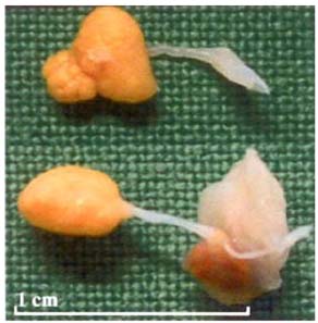
Fig. 1 Macroscopic picture of the rice bodies. The shape and size of those structures vary slightly but both are pedunculated, attached by a thin white strand and composed of a nodular yellow tinted body.
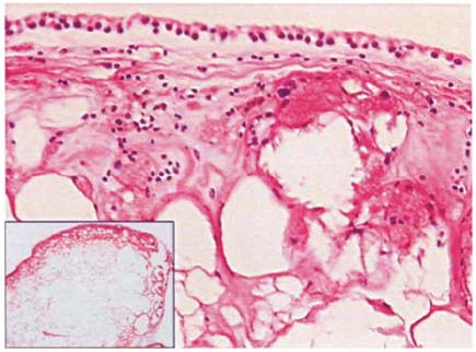
Fig. 2 The rice bodies is mostly composed of benign necrotic adipose tissue. Mild fibrosis, edema and histiocytic infiltration may be seen underneath the synoviocyte lining. H&E stain, ×200. Inset, ×10.
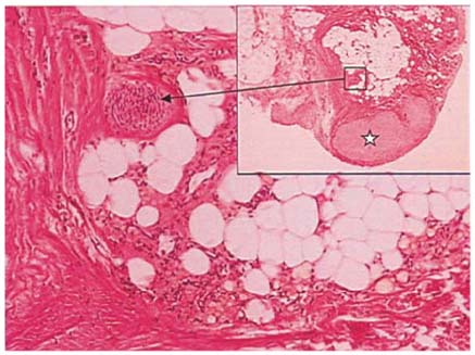
Fig. 3 Picture of the synovial membrane at the base of the second body. Observe the nodular cartilaginous metaplasia (star), and the thickened arteries (arrow). H&E stain, ×100. Inset, ×10.
Reference
-
1. Asik M, Eralp L, Cetik Ö, Altinel L. Rice bodies of synovial origin in the knee joint. Arthroscopy. 2001. 17:E:19.2. Bonnet CS, Walsh DA. Osteoarthritis, angiogenesis and inflammation. Rheumatology (Oxford). 2005. 44:7–16.
Article3. De Bari C, Dell'Accio F, Tylzanowski P, Luyten FP. Multipotent mesenchymal stem cells from adult human synovial membrane. Arthritis Rheum. 2001. 44:1928–1942.
Article4. Gálvez J, Sola J, Ortuño G, Vicente J, Mesa-del Castillo J, Vicente V, Castellon P. Microscopic Rice Bodies in Rheumatoid Synovial Fluid Sediments. J Rheumatol. 1992. 19:1851–1858.5. Geiler G, Mehlhorn U. Vasculitis with anemia infarcts of the villi of the synovial membrane in rheumatoid arthrits. Z Rheumatol. 1989. 48:63–67.6. Peloschek PL. Computergestützte radiologische Quantifizierung der rheumatoiden Arthritis. 1999. University of Vienna;Ph.D. Dissertation.7. Remberger K. Eder M, Gedigk P, editors. Gelenke, Bursen, Sehnenscheiden und Menisci. Allgemeine Pathologie und Pathologische Anatomie. 1990. Berlin: Springer-Verlag;846–866.
- Full Text Links
-
- Actions
-
Cited
- CITED
-
- Close
- Share
-
- Similar articles
-
- A Case of Multiple Rice Bodies by the Nonspecific Synovitis in the Knee Joint
- Multiple Rice Bodies in Subacromial Space: A Case Report
- Rice Bodies Presenting as Intra-Articular Masses in Pediatric Idiopathic Arthritis: A Case Report
- Osteochondral allograft transplantation for treating medial femoral condyle subchondral bone cyst in a 14-year-old standardbred horse: a case report
- Subacromial Bursitis with Rice Bodies: A Case Report
