Characterization of Renal Cell Carcinoma Using Agent Detection Imaging: Comparison with Gray-Scale US
- Affiliations
-
- 1Department of Radiology, Seoul National University College of Medicine, Institute of Radiation Medicine, SNUMRC, and the Clinical Research Institute, Seoul National University Hospital, Korea. kimsh@radcom.snu.ac.kr
- 2Department of Radiology, Samsung Medical Center, Sungkyunkwan University School of Medicine, Korea.
- KMID: 1102714
- DOI: http://doi.org/10.3348/kjr.2005.6.3.173
Abstract
OBJECTIVE
We wanted to compare the imaging features of renal cell carcinoma (RCC) and their diagnostic accuracy on agent detection imaging (ADI) and gray-scale ultrasonography (US). MATERIALS AND METHODS: Thirty non-consecutive patients (age range; 32-80 years, mean age; 53.7 years) with 30 RCCs were examined with gray-scale US and with ADI in conjunction with using SH U 508A. We described the imaging features of the renal tumors obtained from ADI according to their enhancement pattern, the intratumoral anechoic areas and the presence of any pseudocapsule. The imaging features and diagnostic accuracy of ADI and gray-scale US were then compared. RESULTS: On the ADI exam, the RCCs were shown as being heterogeneous in 87% of the cases (26/30), homogeneous in 7% of the cases (2/30), and there was peripheral irregular enhancement in 7% of the cases (2/30). Intratumoral anechoic areas and pseudocapsule were seen in 87% and 77% of the RCCs on the ADI exam, whereas these features were seen in 53% and 17% of the cases on the gray-scale US, respectively. The diagnostic sensitivity, specificity, and accuracy for RCC with ADI were 97%, 93%, and 95%, respectively. However, those for RCC with using gray-scale US were 70%, 86%, and 78%, respectively. There was a significant difference for the diagnostic accuracy of RCC between ADI and gray-scale US (p < 0.05). CONCLUSION: Agent detection imaging can help visualize the enhancement pattern of RCC and improve the diagnostic accuracy of this tumor by better displaying the intratumoral anechoic areas and the pseudocapsule than does the gray-scale US.
Keyword
MeSH Terms
Figure
-
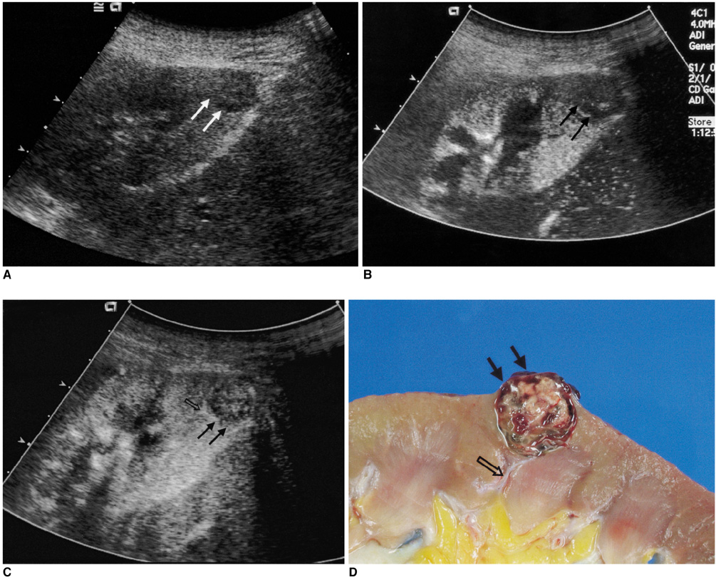
Fig. 1 Small renal cell carcinoma in a 64-year-old man. A. Transverse image on gray-scale US shows an ill-defined isoechoic mass (arrows) containing suspicious anechoic cysts. Any pseudocapsule is not seen around the tumor. B. The transverse image of agent detection imaging that was obtained 17 seconds after the administration of SH U 508A shows a thick pseudocapsule (arrows) around the tumor. C. The transverse image of agent detection imaging that was obtained 34 seconds after the administration of SH U 508A shows heterogeneous enhancement of the tumor with multiple anechoic cysts, and these are better depicted here than on gray-scale US. The pseudocapsule as well as the tumor is also enhanced (arrows). A feeding vessel (blank arrow) is clearly seen on the agent detection imaging exam. D. The cut surface of the gross specimen shows small cysts, necrosis and hemorrhage within the renal cell carcinoma (arrows). A feeding vessel (blank arrow) is demonstrated on the pathologic exam, and this correlated very well with the findings on the agent detection imaging exam.

Fig. 2 Renal cell carcinoma in a 60-year-old man. A. Transverse image of the gray-scale US shows a slightly hyperechoic solid mass (arrows) with no intratumoral anechoic areas and a pseudocapsule in the right kidney. B. Transverse image of agent detection imaging that was obtained 15 seconds after the administration of SH U 508A shows heterogeneous enhancement of the tumor with a thin pseudocapsule (arrows). C. Transverse image of agent detection imaging that was obtained 1 minute after the administration of SH U 508A shows heterogeneous enhancement of the tumor with multiple intratumoral anechoic areas (blank arrows). The pseudocapsule is not clearly seen due to the delayed enhancement. D. The cut surface of the gross specimen shows multiple small cystic and necrotic portions (blank arrows) in the renal cell carcinoma with a pseudocapsule (arrows).
Reference
-
1. Smith SJ, Bosniak MA, Megibow AJ, Hulnick DH, Horii SC, Raghavendra BN. Renal cell carcinoma: earlier discovery and increased detection. Radiology. 1989. 170:699–703.2. Siegel CL, Middleton WD, Teefey SA, McClennan BL. Angiomyolipoma and renal cell carcinoma: US differentiation. Radiology. 1996. 198:789–793.3. Yamashita Y, Ueno S, Makita O, Ogata I, Hatanaka Y, Watanabe O, et al. Hyperechoic renal tumors: anechoic rim and intratumoral cysts in US differentiation of renal cell carcinoma from angiomyolipoma. Radiology. 1993. 188:179–182.4. Jinzaki M, Ohkuma K, Tanimoto A, Mukai M, Hiramatsu K, Murai M, et al. Small solid renal lesions: usefulness of power Doppler US. Radiology. 1998. 209:543–550.5. Kim AY, Kim SH, Kim YJ, Lee IH. Contrast-enhanced power Doppler sonography for the differentiation of cystic renal lesions: preliminary study. J Ultrasound Med. 1999. 18:581–588.6. Ascenti G, Zimbaro G, Mazziotti S, Visalli C, Racchiusa S, Vinci S, et al. Doppler power with contrast media in the characterization of renal masses. Radiol Med (Torino). 2000. 100:168–174.7. Kim TK, Choi BI, Han JK, Hong HS, Park SH, Moon SG. Hepatic tumors: contrast agent-enhancement patterns with pulse-inversion harmonic US. Radiology. 2000. 216:411–417.8. Youk JH, Kim CS, Lee JM. Contrast-enhanced agent detection imaging: value in the characterization of focal hepatic lesions. J Ultrasound Med. 2003. 22:897–910.9. Youk JH, Lee JM, Kim CS. Therapeutic response evaluation of malignant hepatic masses treated by interventional procedures with contrast-enhanced agent detection imaging. J Ultrasound Med. 2003. 22:911–920.10. Quaia E, Siracusano S, Bertolotto M, Monduzzi M, Mucelli RP. Characterization of renal tumours with pulse inversion harmonic imaging by intermittent high mechanical index technique: initial results. Eur Radiol. 2003. 13:1402–1412.11. Kim JH, Eun HW, Lee HK, Park SJ, Shin JH, Hwang JH, et al. Renal perfusion abnormality. coded harmonic angio US with contrast agent. Acta Radiol. 2003. 44:166–171.12. Ascenti G, Gaeta M, Magno C, Mazziotti S, Blandino A, Melloni D, et al. Contrast-enhanced second-harmonic sonography in the detection of pseudocapsule in renal cell carcinoma. AJR Am J Roentgenol. 2004. 182:1525–1530.13. Pickhardt PJ, Lonergan GJ, Davis CJw Jr, Kashitani N, Wagner BJ. From the archives of the AFIP. Infiltrative renal lesions: radiologic-pathologic correlation. Armed Forces Institute of Pathology. Radiographics. 2000. 20:215–243.14. Yamashita Y, Honda S, Nishiharu T, Urata J, Takahashi M. Detection of pseudocapsule of renal cell carcinoma with MR imaging and CT. AJR Am J Roentgenol. 1996. 166:1151–1155.15. Pretorius ES, Siegelman ES, Ramchandani P, Cangiano T, Banner MP. Renal neoplasms amenable to partial nephrectomy: MR imaging. Radiology. 1999. 212:28–34.
- Full Text Links
-
- Actions
-
Cited
- CITED
-
- Close
- Share
-
- Similar articles
-
- Urine Cytology of Renal Cell Carcinoma: Analysis of 11 cases
- Multiphasic spiral CT of renal masses: Comparison among phases following contrast injection
- A study on the comparision of various imaging methods for the staging of renal cell carcinoma
- RE: Distinguishing between Renal Cell Carcinoma and Fat Poor Angiomyolipoma in Diffusion-Weighted Imaging
- A Case of Renal Cell Carcinoma in Childhood
