Korean J Radiol.
2010 Oct;11(5):547-552. 10.3348/kjr.2010.11.5.547.
Estimation of Radiation Exposure of 128-Slice 4D-Perfusion CT for the Assessment of Tumor Vascularity
- Affiliations
-
- 1Department of Diagnostic and Interventional Radiology, University Hospital Tuebingen, Hoppe-Seyler-Strasse 3, 72076 Tuebingen, Germany. dominik.ketelsen@med.uni-tuebingen.de
- 2Departments of Radiotherapy and Radiooncology, University Hospital Tuebingen, Hoppe-Seyler-Strasse 3, 72076 Tuebingen, Germany.
- KMID: 1102580
- DOI: http://doi.org/10.3348/kjr.2010.11.5.547
Abstract
OBJECTIVE
We aimed to estimate the effective dose of 4D-Perfusion-CT protocols of the lung, liver, and pelvis for the assessment of tumor vascularity.
MATERIALS AND METHODS
An Alderson-Rando phantom equipped with thermoluminescent dosimeters was used to determine the effective dose values of 4D-Perfusion-CT. Phantom measurements were performed on a 128-slice single-source scanner in adaptive 4D-spiral-mode with bidirectional table movement and a total scan range of 69 mm over a time period of nearly 120 seconds (26 scans). Perfusion measurements were simulated for the lung, liver, and pelvis under the following conditions: lung (80 kV, 60 mAs), liver (80 kV/80 mAs and 80 kV/120 mAs), pelvis (100 kV/80 mAs and 100 kV/120 mAs).
RESULTS
Depending on gender, the evaluated body region and scan protocol, an effective whole-body dose between 2.9-12.2 mSv, was determined. The radiation exposure administered to gender-specific organs like the female breast tissue (lung perfusion) or to the ovaries (pelvic perfusion) led to an increase in the female specific dose by 86% and 100% in perfusion scans of the lung and the pelvis, respectively.
CONCLUSION
Due to a significant radiation dose of 4D-perfusion-CT protocols, the responsible use of this new promising technique is mandatory. Gender- and organ-specific differences should be considered for indication and planning of tumor perfusion scans.
Keyword
MeSH Terms
Figure
-
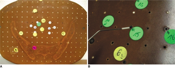
Fig. 1 Dosimeters and dosimetry.A, B. One 25 mm slice of 35 layers (A) of Alderson-Rando phantom at pelvis, with numbering of borehole positions representing pelvic organs. TLD is positioned to defined borehole with aid of vacuum forceps (B).

Fig. 2 Gender-specific estimated effective whole-body dose of different 4D perfusion protocols with scan range of 69 mm for normal weighted patients.
Reference
-
1. Miles KA. Perfusion CT for the assessment of tumour vascularity: which protocol? Br J Radiol. 2003; 76:S36–S42. PMID: 15456712.
Article2. Wu GY, Ghimire P. Perfusion computed tomography in colorectal cancer: protocols, clinical applications and emerging trends. World J Gastroenterol. 2009; 15:3228–3231. PMID: 19598297.
Article3. Li WW. Tumor angiogenesis: molecular pathology, therapeutic targeting, and imaging. Acad Radiol. 2000; 7:800–811. PMID: 11048878.
Article4. Ketelsen D, Thomas C, Werner M, Luetkhoff MH, Buchgeister M, Tsiflikas I, et al. Dual-source computed tomography: estimation of radiation exposure of ECG-gated and ECG-triggered coronary angiography. Eur J Radiol. 2010; 73:274–279. PMID: 19097836.
Article5. Hunold P, Vogt FM, Schmermund A, Debatin JF, Kerkhoff G, Budde T, et al. Radiation exposure during cardiac CT: effective doses at multi-detector row CT and electron-beam CT. Radiology. 2003; 226:145–152. PMID: 12511683.
Article6. Ketelsen D, Luetkhoff MH, Thomas C, Werner M, Buchgeister M, Tsiflikas I, et al. Estimation of the radiation exposure of a chest pain protocol with ECG-gating in dual-source computed tomography. Eur Radiol. 2009; 19:37–41. PMID: 18648818.
Article7. The 2007 Recommendations of the International Commission on Radiological Protection. ICRP publication 103. Ann ICRP. 2007; 37:1–332.8. Ng QS, Goh V, Klotz E, Fichte H, Saunders MI, Hoskin PJ, et al. Quantitative assessment of lung cancer perfusion using MDCT: does measurement reproducibility improve with greater tumor volume coverage? AJR Am J Roentgenol. 2006; 187:1079–1084. PMID: 16985160.
Article9. Park MS, Klotz E, Kim MJ, Song SY, Park SW, Cha SW, et al. Perfusion CT: noninvasive surrogate marker for stratification of pancreatic cancer response to concurrent chemo- and radiation therapy. Radiology. 2009; 250:110–117. PMID: 18984781.
Article10. Ng CS, Wang X, Faria SC, Lin E, Charnsangavej C, Tannir NM. Perfusion CT in patients with metastatic renal cell carcinoma treated with interferon. AJR Am J Roentgenol. 2010; 194:166–171. PMID: 20028919.
Article11. Petralia G, Preda L, D'Andrea G, Viotti S, Bonello L, De Filippi R, et al. CT perfusion in solid-body tumours. Part I: technical issues. Radiol Med. 2010; [Epub ahead of print].
Article12. Yang HF, Du Y, Ni JX, Zhou XP, Li JD, Zhang Q, et al. Perfusion computed tomography evaluation of angiogenesis in liver cancer. Eur Radiol. 2010; 20:1424–1430. PMID: 20179942.
Article13. Bellomi M, Viotti S, Preda L, D'Andrea G, Bonello L, Petralia G. Perfusion CT in solid body-tumours part II. Clinical applications and future development. Radiol Med. 2010; [Epub ahead of print].
Article14. Zhong L, Wang WJ, Xu JR. Clinical application of hepatic CT perfusion. World J Gastroenterol. 2009; 15:907–911. PMID: 19248188.
Article15. Bellomi M, Petralia G, Sonzogni A, Zampino MG, Rocca A. CT perfusion for the monitoring of neoadjuvant chemotherapy and radiation therapy in rectal carcinoma: initial experience. Radiology. 2007; 244:486–493. PMID: 17641369.
Article16. Cuenod CA, Fournier L, Balvay D, Guinebretière JM. Tumor angiogenesis: pathophysiology and implications for contrast-enhanced MRI and CT assessment. Abdom Imaging. 2006; 31:188–193. PMID: 16447089.
Article17. Kapanen M, Halavaara J, Häkkinen AM. Comparison of liver perfusion parameters studied with conventional extravascular and experimental intravascular CT contrast agents. Acad Radiol. 2007; 14:951–958. PMID: 17659241.
Article18. Kudo M. Imaging blood flow characteristics of hepatocellular carcinoma. Oncology. 2002; 62:48–56. PMID: 11868785.
Article19. Petralia G, Preda L, Giugliano G, Jereczek-Fossa BA, Rocca A, D'Andrea G, et al. Perfusion computed tomography for monitoring induction chemotherapy in patients with squamous cell carcinoma of the upper aerodigestive tract: correlation between changes in tumor perfusion and tumor volume. J Comput Assist Tomogr. 2009; 33:552–559. PMID: 19638848.20. Gillard JH, Antoun NM, Burnet NG, Pickard JD. Reproducibility of quantitative CT perfusion imaging. Br J Radiol. 2001; 74:552–555. PMID: 11459735.
Article21. Brenner DJ, Hall EJ. Computed tomography--an increasing source of radiation exposure. N Engl J Med. 2007; 357:2277–2284. PMID: 18046031.22. Christner JA, Kofler JM, McCollough CH. Estimating effective dose for CT using dose-length product compared with using organ doses: consequences of adopting International Commission on Radiological Protection publication 103 or dual-energy scanning. AJR Am J Roentgenol. 2010; 194:881–889. PMID: 20308486.
Article23. Boetticher H, Lachmund J, Looe HK, Hoffmann W, Poppe B. 2007 recommendations of the ICRP change basis for estimation of the effective dose: what is the impact on radiation dose assessment of patient and personnel? Rofo. 2008; 180:391–395. PMID: 18438741.
Article24. Prakash P, Kalra MK, Gilman MD, Shepard JA, Digumarthy SR. Is weight-based adjustment of automatic exposure control necessary for the reduction of chest CT radiation dose? Korean J Radiol. 2010; 11:46–53. PMID: 20046494.
Article25. Kalra MK, Dang P, Singh S, Saini S, Shepard JA. In-plane shielding for CT: effect of off-centering, automatic exposure control and shield-to-surface distance. Korean J Radiol. 2009; 10:156–163. PMID: 19270862.
Article
- Full Text Links
-
- Actions
-
Cited
- CITED
-
- Close
- Share
-
- Similar articles
-
- A study on radiation exposure dose at brain CT
- Strategy of Respiratory Gated Radiation Therapy for Lung Cancer Patient in Stereotactic Radiosurgery : Phantom Study
- Four-Dimensional Thoracic CT in Free-Breathing Children
- Pediatric CT: Understanding of Radiation Dose and Optimization of Imaging Techniques
- Assessment of Respiratory Tumor Movement using 4D Computed Tomography for Stereotactic Radiosurgery in Lung Tumor
