Ultrasonographic Features of Benign Adenomyoepithelioma of the Breast
- Affiliations
-
- 1Department of Radiology, Seoul St. Mary's Hospital, The Catholic University of Korea, Seoul 137-701, Korea. rad-ksh@catholic.ac.kr
- 2Department of Pathology, Seoul St. Mary's Hospital, The Catholic University of Korea, Seoul 137-701, Korea.
- 3Department of Surgery, Seoul St. Mary's Hospital, The Catholic University of Korea, Seoul 137-701, Korea.
- KMID: 1102576
- DOI: http://doi.org/10.3348/kjr.2010.11.5.522
Abstract
OBJECTIVE
The purpose of this study was to evaluate the ultrasonographic features of benign adenomyoepithelioma of the breast.
MATERIALS AND METHODS
Between 2005 and 2009, five patients had histologically confirmed adenomyoepithelioma of the breast. We retrospectively evaluated the ultrasonographic findings of the tumors in correlation with the pathology, and reviewed medical records.
RESULTS
The clinical manifestations included a palpable mass in three patients, while mammographic screening helped detect abnormalities in two patients. Ultrasonograms showed masses with an oval (n = 3) or irregular (n = 2) shape, with uncircumscribed (n = 4) or relatively well-circumscribed (n = 1) margins, as well as with a hypoechoic (n = 3) or a complex echoic (n = 2) internal echo texture. Three patients had focal ductectasia adjacent to the mass. The ultrasonographic assessments were classified as Breast Imaging Reporting and Data System (BI-RADS) category 4A, with low suspicion of malignancy in two cases, and as category 4B, with intermediate suspicion of malignancy in three cases. The pathology revealed benign adenomyoepithelioma in all patients.
CONCLUSION
Benign adenomyoepitheliomas appear as solid or complex echoic masses with suspicious malignant ultrasonographic features, which may be associated with adjacent ductectasia. Although adenomyoepithelioma is a rare breast tumor, awareness of its sonographic features will be helpful for the differential diagnosis from other tumors.
Keyword
MeSH Terms
Figure
-
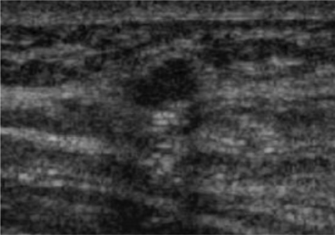
Fig. 1 72-year-old woman with no symptoms (Patient 1). Ultrasonogram revealed oval microlobulated hypoechoic mass. BI-RADS assessment was classified as category 4A, with low suspicion of malignancy.
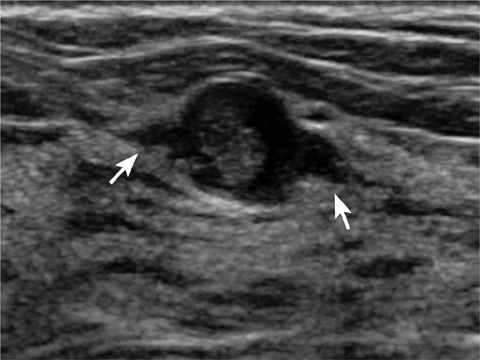
Fig. 2 38-year-old woman with no symptoms (Patient 2). Ultrasonogram revealed oval circumscribed mass with complex echogenicity and adjacent duct dilatations (arrows). BI-RADS assessment was classified as category 4A.

Fig. 3 31-year-old woman with palpable mass (Patient 3). A. Ultrasonogram revealed oval hypoechoic mass with angular margin and adjacent focal duct dilatations (arrows). BI-RADS assessment was classified as category 4B. B. Craniocaudal mammogram showed oval obscured isodense mass (arrows) without calcification. C. Photomicrograph of histologic specimen from excision revealed well-circumscribed adenomyoepithelioma with adjacent dilatated ducts (*) (Hematoxylin & Eosin staining, ×40). D. Adenomyoepithelioma with gland and cord-like growth pattern revealed prominent myoepithelial cell hyperplasia. Focal apocrine metaplasia of epithelial cells are noted (arrows) (Hematoxylin & Eosin staining, ×200).
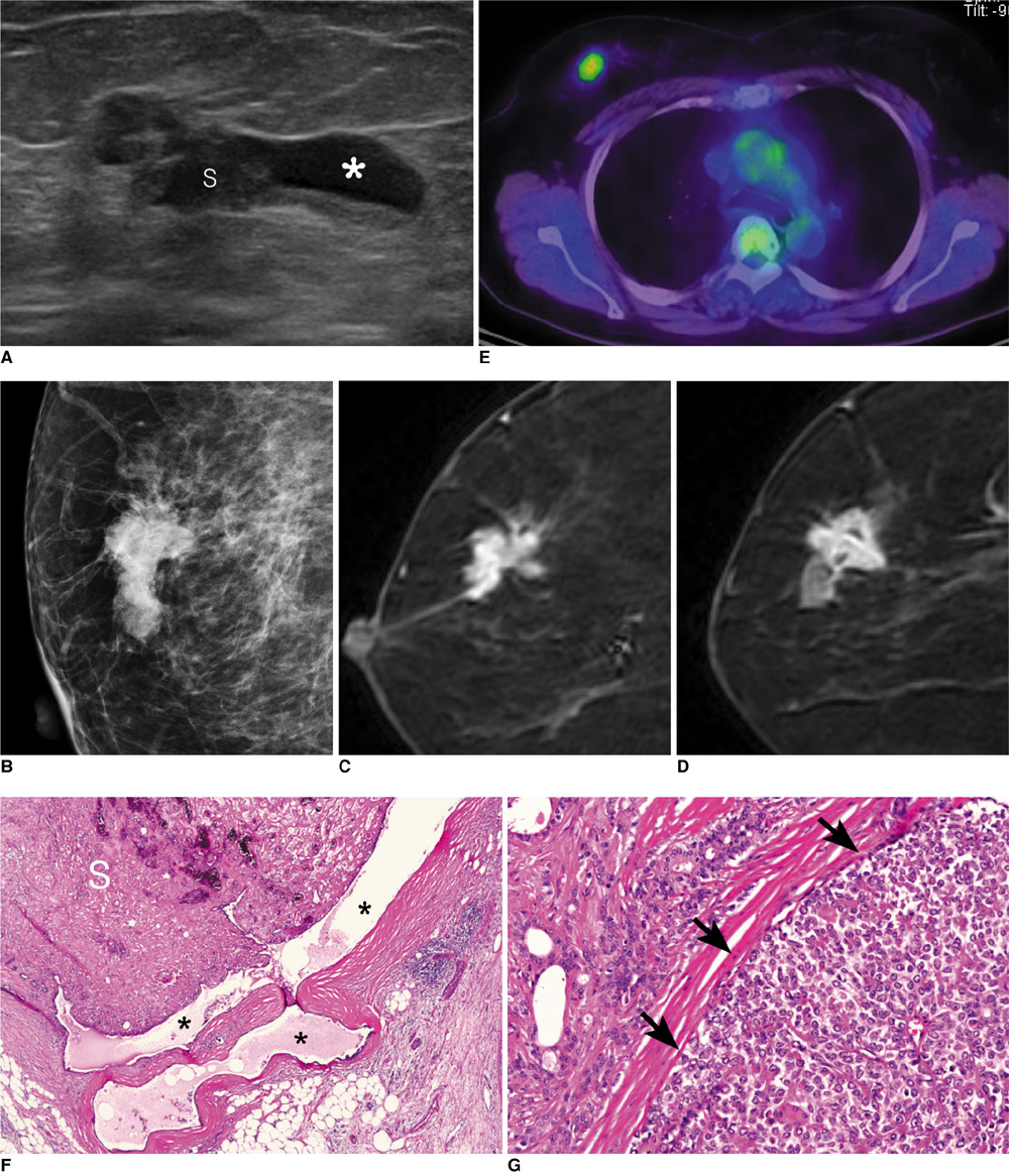
Fig. 4 54-year-old woman with palpable mass (Patient 5). A. Ultrasonogram revealed irregular, angular, and complex echoic mass with solid (S) and cystic (*) portions. BI-RADS assessment was classified as category 4B. B. Mediolateral oblique mammogram revealed irregular, indistinct hyperdense mass with linear microcalcifications. C, D. MRI revealed irregular, spiculated mass with heterogeneous enhancement and cystic portions. E. PET/CT revealed FDG uptake with 4.9 SUVmax at correlating mass. F. Photomicrograph of histologic specimen from excision revealed adenomyoepithelioma with solid (S) and cystic (*) components (Hematoxylin & Eosin staining, ×40). G. Focal atypical proliferation of myoepithelial cells (arrows) in otherwise typical adenomyoepithelioma (Hematoxylin & Eosin staining, ×200).
Cited by 1 articles
-
Rapid Local Recurrence of Breast Myoepithelial Carcinoma Arising in Adenomyoepithelioma: A Case Report
Mo In Ha, Bo Kyoung Seo, Jung Woo Choi
J Korean Soc Radiol. 2020;81(1):207-212. doi: 10.3348/jksr.2020.81.1.207.
Reference
-
1. Hamperl H. The myothelia (myoepithelial cells). Normal state; regressive changes; hyperplasia; tumors. Curr Top Pathol. 1970. 53:161–220.2. Rosen PP. Adenomyoepithelioma of the breast. Hum Pathol. 1987. 18:1232–1237.3. Loose JH, Patchefsky AS, Hollander IJ, Lavin LS, Cooper HS, Katz SM. Adenomyoepithelioma of the breast. A spectrum of biologic behavior. Am J Surg Pathol. 1992. 16:868–876.4. Tavassoli FA. Myoepithelial lesions of the breast. Myoepitheliosis, adenomyoepithelioma, and myoepithelial carcinoma. Am J Surg Pathol. 1991. 15:554–568.5. Howlett DC, Mason CH, Biswas S, Sangle PD, Rubin G, Allan SM. Adenomyoepithelioma of the breast: spectrum of disease with associated imaging and pathology. AJR Am J Roentgenol. 2003. 180:799–803.6. Fan F, Smith W, Wang X, Jewell W, Thomas PA, Tawfik O. Myoepithelial carcinoma of the breast arising in an adenomyoepithelioma: mammographic, ultrasound and histologic features. Breast J. 2007. 13:203–204.7. Felipo F, Del Agua C, Eguizabal C, Vaquero M. Benign adenomyoepithelioma of the breast: a case with gross mimicking of malignancy. Breast J. 2002. 8:383–384.8. Tukel S, Ustuner E, Aytac SK. Adenomyoepithelioma of the breast. J Ultrasound Med. 2001. 20:1021–1024.9. Ruiz-Delgado ML, Lopez-Ruiz JA, Eizaguirre B, Saiz A, Astigarraga E, Fernandez-Temprano Z. Benign adenomyoepithelioma of the breast: imaging findings mimicking malignancy and histopathological features. Acta Radiol. 2007. 48:27–29.10. Ahmed AA, Heller DS. Malignant adenomyoepithelioma of the breast with malignant proliferation of epithelial and myoepithelial elements: a case report and review of the literature. Arch Pathol Lab Med. 2000. 124:632–636.11. Lee WY. Fine needle aspiration cytology of adenomyoepithelioma of the breast: a case indistinguishable from phyllodes tumor in cytologic findings and clinical behavior. Acta Cytol. 2000. 44:488–490.12. Hikino H, Kodama K, Yasui K, Ozaki N, Nagaoka S, Miura H. Intracystic adenomyoepithelioma of the breast--case report and review. Breast Cancer. 2007. 14:429–433.13. Papaevangelou A, Pougouras I, Liapi G, Pierrakakis S, Tibishrani M, Setakis N. Cystic adenomyoepithelioma of the breast. Breast. 2004. 13:356–358.14. Noël JC, Simon P, Aguilar SF. Malignant myoepithelioma arising in cystic adenomyoepithelioma. Breast J. 2006. 12:386.
