Pulmonary Complication of Novel Influenza A (H1N1) Infection: Imaging Features in Two Patients
- Affiliations
-
- 1Department of Radiology and Research Institute of Radiology, Asan Medical Center, University of Ulsan College of Medicine, Seoul 138-736, Korea. seojb@amc.seoul.kr
- 2Department of Respiratory and Critical Care Medicine, Asan Medical Center, University of Ulsan College of Medicine, Seoul 138-736, Korea.
- 3Department of Emergency Medicine, Asan Medical Center, University of Ulsan College of Medicine, Seoul 138-736, Korea.
- KMID: 1102555
- DOI: http://doi.org/10.3348/kjr.2009.10.6.531
Abstract
- Novel influenza A (H1N1) virus is the pathogen of recent global outbreaks of febrile respiratory infection. We herein report the imaging findings of pulmonary complication in two patients with novel influenza A (H1N1) infection. The first patient without secondary infection showed the ill-defined ground-glass opacity nodules and patch areas of ground-glass opacities. The second patient with secondary pneumococcal pneumonia showed areas of lobar consolidation in the right middle lobe and left lower lobe and ground-glass opacities.
Keyword
MeSH Terms
Figure
-

Fig. 1 16-year-old girl diagnosed as novel influenza A (H1N1) pneumonia without secondary infection. A. Initial chest radiograph shows ill-defined increased opacity in right lower lung zone. B, C. High-resolution chest CT scans show ill-defined ground-glass opacities with interlobular septal thickening and some ill-defined nodules in right middle and lower lobes. D. Follow-up chest radiograph after medication shows improvement of infiltration in lung.
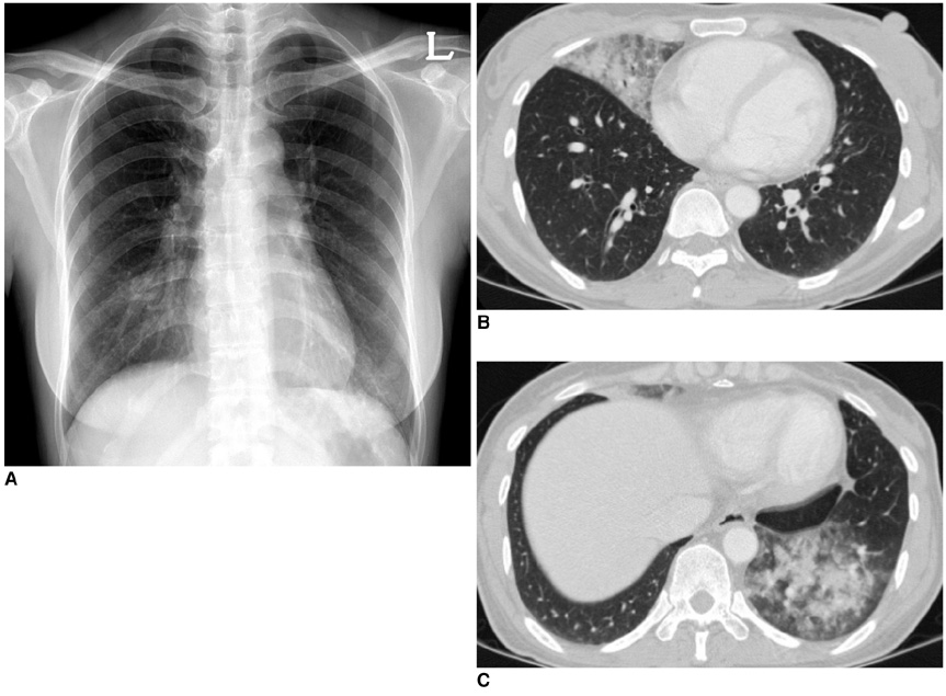
Fig. 2 42-year-old woman diagnosed as novel influenza A (H1N1) pneumonia with secondary pneumococcal pneumonia. A. Initial chest radiograph shows ill-defined infiltrates in both lower lung zones. B, C. Chest CT scans show lobar-distributed ill-defined consolidation and peripheral ground-glass opacities in right middle lobe and left lower lobe.
Cited by 1 articles
-
Comparison of Clinical Manifestation and Laboratory Findings between H1N1 and Influenza B Infection
Su Hee Kim, Chul Hyue Park, Kyoung Huh, Gyu Hong Shim, Hyo-Bin Kim, Su Jeong You, Young Whan Song, Ju-Young Chung, Mi Jung Park, Chang-Keun Kim, Myoung Jae Chey, Ja Wook Koo, Sang Woo Kim
Pediatr Allergy Respir Dis. 2012;22(1):64-70. doi: 10.7581/pard.2012.22.1.64.
Reference
-
1. Dawood FS, Jain S, Finelli L, Shaw MW, Lindstrom S, Garten RJ, et al. Emergence of a novel swine-origin influenza a (H1N1) virus in humans. N Engl J Med. 2009. 360:2605–2615.2. Perez-Padilla R, de la Rosa-Zamboni D, Ponce de Leon S, Hernandez M, Quinones-Falconi F, Bautista E, et al. Pneumonia and respiratory failure from swine-origin influenza a (H1N1) in Mexico. N Engl J Med. 2009. 361:680–689.3. Chowell G, Bertozzi SM, Colchero MA, Lopez-Gatell H, Alpuche-Aranda C, Hernandez M, et al. Severe respiratory disease concurrent with the circulation of H1N1 influenza. N Engl J Med. 2009. 361:674–679.4. Hancock K, Veguilla V, Lu X, Zhong W, Butler EN, Sun H, et al. Cross-reactive antibody responses to the 2009 pandemic H1N1 influenza virus. N Engl J Med. 2009. [Epub ahead of print].5. Jordan H, Mosquera M, Nair H, France A. [ME1]Swine-origin influenza a (H1N1) virus infections in a school - New York City, April 2009. MMWR Morb Mortal Wkly Rep. 2009. 58:470–472.6. Fiore AE, Shay DK, Broder K, Iskander JK, Uyeki TM, Mootrey G, et al. Prevention and control of influenza: recommendations of the Advisory Committee on Immunization Practices (ACIP), 2008. MMWR Recomm Rep. 2008. 57:1–60.7. Kim EA, Lee KS, Primack SL, Yoon HK, Byun HS, Kim TS, et al. Viral pneumonias in adults: radiologic and pathologic findings. Radiographics. 2002. 22:S137–S149.
- Full Text Links
-
- Actions
-
Cited
- CITED
-
- Close
- Share
-
- Similar articles
-
- Encephalitis Induced by 2009 H1N1 Influenza A
- Influenza Associated Pneumonia
- Pandemic Influenza (H1N1) and Mycobacterium tuberculosis Co-infection
- A Case of Novel Influenza A (H1N1) Virus Pneumonia Complicated Pnemomediastinum and Subcutenous Emphysema
- Epidemiology, clinical manifestations, and management of pandemic novel Influenza A (H1N1)
