A Comparison of the Use of Contrast Media with Different Iodine Concentrations for Multidetector CT of the Kidney
- Affiliations
-
- 1Department of Radiology, Seoul National University College of Medicine, Seoul, Korea, Institute of Radiation Medicine and Kidney Research Institute, Seoul National University Medical Research Center, Seoul 110-744, Korea. kimsh@radcom.snu.ac.kr
- KMID: 1101926
- DOI: http://doi.org/10.3348/kjr.2011.12.6.714
Abstract
OBJECTIVE
To determine the optimal iodine concentration of contrast media for kidney multidetector computed tomography (MDCT) by comparing the degree of renal parenchymal enhancement and the severity of the renal streak artifact with contrast media of different iodine concentrations.
MATERIALS AND METHODS
A 16-row MDCT was performed in 15 sedated rabbits by injection of 2 mL contrast media/kg body weight at a rate of 0.3 mL/sec. Monomeric nonionic contrast media of 250, 300, and 370 mg iodine/mL were injected at 1-week intervals. Mean attenuation values were measured in each renal structure with attenuation differences among the structures. The artifact was evaluated by CT window width/level and three grading methods. The values were compared with iodine concentrations.
RESULTS
The 370 mg iodine/mL concentration showed significantly higher cortical enhancement than 250 mg iodine/mL in all phases (p < 0.05). There was however no significant difference in the degree of enhancement between the 300 mg iodine/mL and 370 mg iodine/mL concentrations in all phases. There is a significant difference in attenuation for the cortex-outer medulla between 250 mg iodine/mL and 300 mg iodine/mL (p < 0.05). The artifact was more severe with a medium of 370 mg iodine/mL than with 250 mg iodine/mL by all grading methods (p < 0.05).
CONCLUSION
The 300 mg iodine/mL is considered to be the most appropriate iodine concentration in an aspect of the enhancement and artifact on a kidney MDCT scan.
Keyword
MeSH Terms
Figure
-
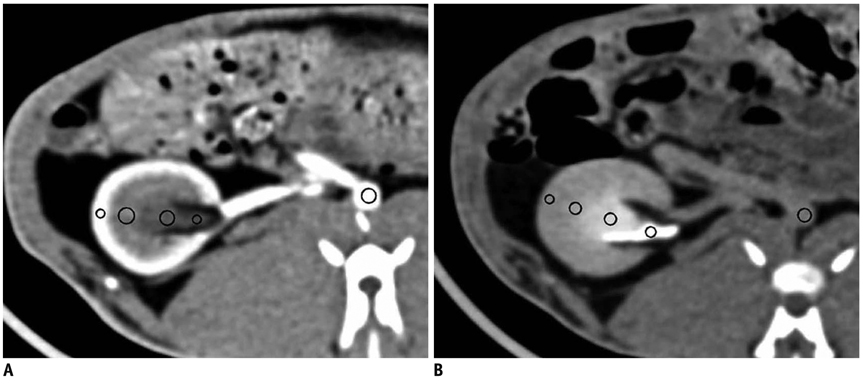
Fig. 1 Axial contrast enhanced CT images show five region-of-interest circles at 5-second delay (A) and 115-second delay (B) for 370 mg iodine/mL. Attenuation values were measured using region-of-interest circles. Region-of-interest circles were located in cortex, outer medulla, inner medulla, pelvis and aorta as depicted on PACS workstation.
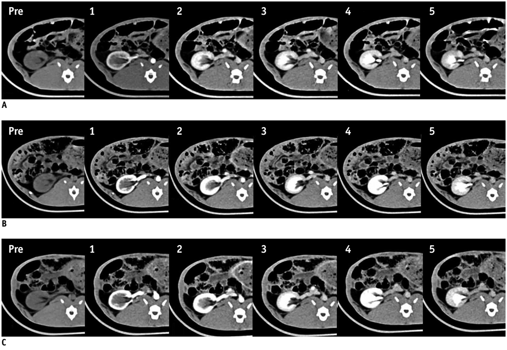
Fig. 2 Axial contrast enhanced CT images in rabbit show various CT enhancement patterns according to six phases and use of contrast media with three iodine concentrations. CT images show prominent differences of enhancement according to iodine concentration of contrast media in renal cortex regardless of phase, with most severe renal steak artifact produced with concentration of 370 mg iodine/mL. Three rows indicate iodine concentrations: A. 250 mg iodine/mL, B. 300 mg iodine/mL, C. 370 mg iodine/mL. Six columns indicate each phase as function of time: pre (pre-contrast), phase 1 (5-second delay), phase 2 (15-second delay), phase 3 (35-second delay), phase 4 (65-second delay), and phase 5 (115-second delay).
Cited by 1 articles
-
Use of Iterative Reconstruction and a Small Contrast Volume in Rabbit Kidney CT: Comparison with Conventional Protocol
Rihyeon Kim, Sang Youn Kim, Jeong Yeon Cho, Joongyub Lee, Seung Hyup Kim
J Korean Soc Radiol. 2018;79(2):77-87. doi: 10.3348/jksr.2018.79.2.77.
Reference
-
1. Marchiano A, Spreafico C, Lanocita R, Frigerio L, Di Tolla G, Patelli G, et al. Does iodine concentration affect the diagnostic efficacy of biphasic spiral CT in patients with hepatocellular carcinoma? Abdom Imaging. 2005. 30:274–280.2. Yamashita Y, Komohara Y, Takahashi M, Uchida M, Hayabuchi N, Shimizu T, et al. Abdominal helical CT: evaluation of optimal doses of intravenous contrast material--a prospective randomized study. Radiology. 2000. 216:718–723.3. Fenchel S, Fleiter TR, Aschoff AJ, van Gessel R, Brambs HJ, Merkle EM. Effect of iodine concentration of contrast media on contrast enhancement in multislice CT of the pancreas. Br J Radiol. 2004. 77:821–830.4. Cademartiri F, Mollet NR, van der Lugt A, McFadden EP, Stijnen T, de Feyter PJ, et al. Intravenous contrast material administration at helical 16-detector row CT coronary angiography: effect of iodine concentration on vascular attenuation. Radiology. 2005. 236:661–665.5. Sandstede JJ, Kaupert C, Roth A, Jenett M, Harz C, Hahn D. Comparison of different iodine concentrations for multidetector row computed tomography angiography of segmental renal arteries. Eur Radiol. 2005. 15:1211–1214.6. Sandstede JJ, Werner A, Kaupert C, Roth A, Jenett M, Harz C, et al. A prospective study comparing different iodine concentrations for triphasic multidetector row CT of the upper abdomen. Eur J Radiol. 2006. 60:95–99.7. Setty BN, Sahani DV, Ouellette-Piazzo K, Hahn PF, Shepard JA. Comparison of enhancement, image quality, cost, and adverse reactions using 2 different contrast medium concentrations for routine chest CT on 16-slice MDCT. J Comput Assist Tomogr. 2006. 30:818–822.8. Bayrak IK, Ozmen Z, Nural MS, Danaci M, Diren B. A comparison of low-dose and normal-dose gadobutrol in MR renography and renal angiography. Korean J Radiol. 2008. 9:250–257.9. Singer AA, Tagliabue JR, Paushter DM, Borkowski GP, Einstein DM. Comparison of iohexol-240 versus iohexol-300 in abdominal CT. Gastrointest Radiol. 1992. 17:122–124.10. Furuta A, Ito K, Fujita T, Koike S, Shimizu A, Matsunaga N. Hepatic enhancement in multiphasic contrast-enhanced MDCT: comparison of high- and low-iodine-concentration contrast medium in same patients with chronic liver disease. AJR Am J Roentgenol. 2004. 183:157–162.11. Behrendt FF, Mahnken AH, Stanzel S, Seidensticker P, Jost E, Gunther RW, et al. Intraindividual comparison of contrast media concentrations for combined abdominal and thoracic MDCT. AJR Am J Roentgenol. 2008. 191:145–150.12. Tozaki M, Naruo K, Fukuda K. Dynamic contrast-enhanced MDCT of the liver: analysis of the effect of different iodine concentrations with the same total iodine dose in the same chronic liver disease patients. Radiat Med. 2005. 23:533–538.13. Bae KT, Heiken JP, Brink JA. Aortic and hepatic contrast medium enhancement at CT. Part I. Prediction with a computer model. Radiology. 1998. 207:647–655.14. Bae KT, Tran HQ, Heiken JP. Uniform vascular contrast enhancement and reduced contrast medium volume achieved by using exponentially decelerated contrast material injection method. Radiology. 2004. 231:732–736.15. Bae KT, Heiken JP, Brink JA. Aortic and hepatic contrast medium enhancement at CT. Part II. Effect of reduced cardiac output in a porcine model. Radiology. 1998. 207:657–662.16. Tello R, Seltzer S. Effects of injection rates of contrast material on arterial phase hepatic CT. AJR Am J Roentgenol. 1999. 173:237–238.17. Sussman SK, Illescas FF, Opalacz JP, Yirga P, Foley LC. Renal streak artifact during contrast-enhanced CT: comparison of low versus high osmolality contrast media. Abdom Imaging. 1993. 18:180–185.18. Tsurusaki M, Sugimoto K, Fujii M, Sugimura K. Multi-detector row helical CT of the liver: quantitative assessment of iodine concentration of intravenous contrast material on multiphasic CT--a prospective randomized study. Radiat Med. 2004. 22:239–245.19. Thomsen HS, Morcos SK, Erley CM, Grazioli L, Bonomo L, Ni Z, et al. The ACTIVE Trial: comparison of the effects on renal function of iomeprol-400 and iodixanol-320 in patients with chronic kidney disease undergoing abdominal computed tomography. Invest Radiol. 2008. 43:170–178.20. Sultana S, Morishita S, Awai K, Kawanaka K, Ohyama Y, Nakayama Y, et al. Evaluation of hypervascular hepatocellular carcinoma in cirrhotic liver by means of helical CT: comparison of different contrast medium concentrations within the same patient. Radiat Med. 2003. 21:239–245.21. Engeroff B, Kopka L, Harz C, Grabbe E. [Impact of different iodine concentrations on abdominal enhancement in biphasic multislice helical CT (MS-CT)]. Rofo. 2001. 173:938–941.22. Fenchel S, Boll DT, Fleiter TR, Brambs HJ, Merkle EM. Multislice helical CT of the pancreas and spleen. Eur J Radiol. 2003. 45:Suppl 1. S59–S72.23. Young SW, Muller HH, Marshall WH. Computed tomography: beam hardening and environmental density artifact. Radiology. 1983. 148:279–283.24. Kuhn MJ, Chen N, Sahani DV, Reimer D, van Beek EJ, Heiken JP, et al. The PREDICT study: a randomized double-blind comparison of contrast-induced nephropathy after low- or isoosmolar contrast agent exposure. AJR Am J Roentgenol. 2008. 191:151–157.
- Full Text Links
-
- Actions
-
Cited
- CITED
-
- Close
- Share
-
- Similar articles
-
- Transient Orbitofacial Angioedema due to Intravenous Iodinated Contrast Media During Computed Tomography: CT Findings
- Analysis of the Effects of Different Iodine Concentrations on the Characterization of Small Renal Lesions Detected by Multidetector Computed Tomography Scan: A Pilot Study
- Comparison of enhancement and image quality: different iodine concentrations for liver on 128-slice multidetector computed tomography in the same chronic liver disease patients
- A study on renal damage in rats induced by different concentrations and osmolarities of diatrizoate
- Technical Aspect of Coronary CT Angiography: Imaging Tips and Safety Issues
