Ultrastructural Changes of Myotendinous Nerve Endings following Recession or Resection Procedures of Extraocular Muscle Surgeries in Cats
- Affiliations
-
- 1Department of Ophthalmology, Korea University College of Medicine, Seoul, Korea. earth317@yahoo.co.kr
- KMID: 1099085
- DOI: http://doi.org/10.3341/kjo.2005.19.1.47
Abstract
- To verify the postoperative ultrastructural changes of the myotendinous nerve endings of feline extraocular muscles, which are known as proprioceptors. Sixteen recti of four cats were used and divided into three groups. In group A, eight lateral recti were recessed. In group B, four medial recti were resected by 10 mm from insertion to include the myotendinous junction. In group C, four medial recti were resected by 4 mm of muscle bellies only, without disturbing the myotendinous junction. Four weeks after surgery, specimens were examined with electron microscopy. In group A, overall neural structures were well maintained with slight axonal degeneration. In group B, only muscle fibers were observed without any regeneration of neural sprouts. In group C, axonal disintegration and shrinkage were evident. These results indicate that myotendinous nerve endings can be damaged in strabismus surgery, and that resection was more invasive than recession in disrupting myotendinous nerve endings.
Keyword
MeSH Terms
Figure
-
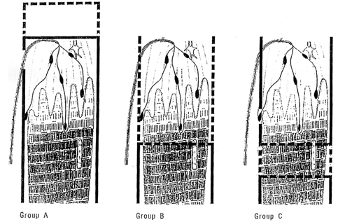
Fig. 1 Schematic drawings of the relationship between operative procedures and palisade endings at the myotendinous junction in each experimental group: A (recession group), B (conventional resection group) and C (modified resection group).
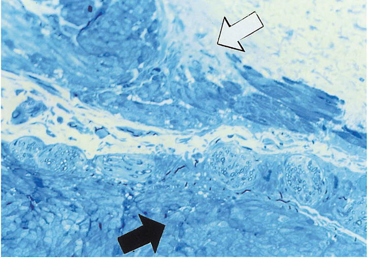
Fig. 2 Cross sectional view of normal nerve endings in the distal myotendinous junction of cat medial rectus. Muscular (black arrow) and tendinous (white arrow) areas are seen (Toluidine blue stain ×400).
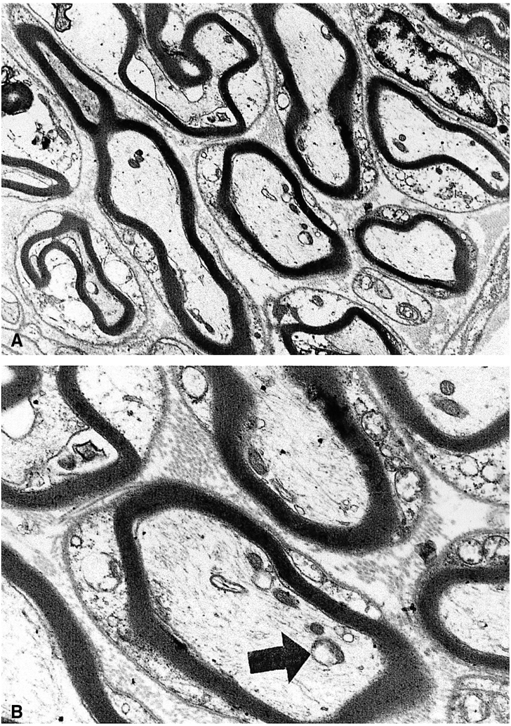
Fig. 3 Cross sectional view of nerve endings at the myotendinous junction of group A. (A) Overall neural structures are well preserved, but microtubules are decreased and neurofilaments increased slightly in the axonal cytoplasm. Axons are well myelinated, but the myelin thickness is decreased slightly (×6000). (B) Mitochondrial swelling is seen (×12,000).

Fig. 4 Cross sectional view of the insertional area of group B. Only muscle fibers are observed without neural structure (×6000).
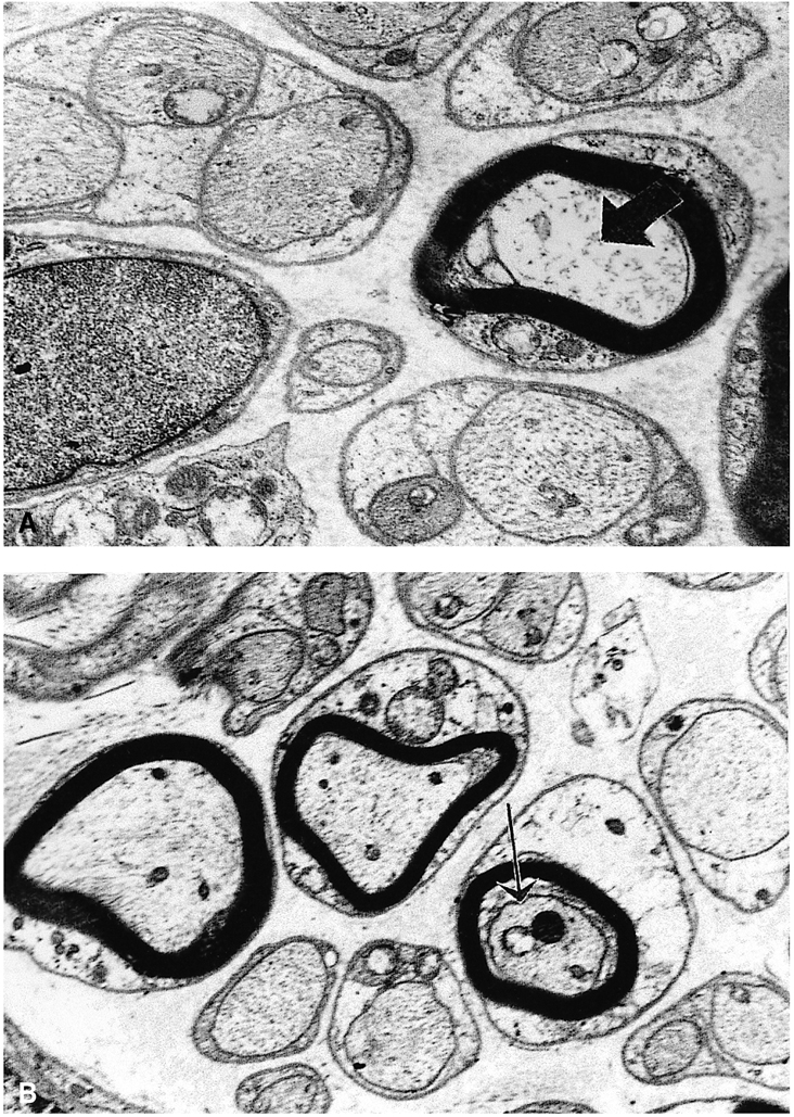
Fig. 5 Cross sectional view of nerve endings at the myotendinous junction of group C. (A) Microtubules are decreased and neurofilaments increased in the axonal cytoplasm. Granular disintegrated axon (black arrow) is also seen (×10,000). (B) Axonal shrinkage as a typical axonal degenerative sign is also seen (thin arrow) (×6,000).
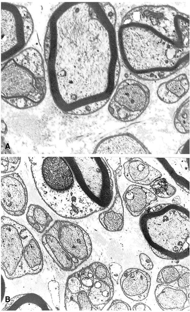
Fig. 6 Cross sectional view of nerve endings at the myotendinous junction of the normal group. The integrity of axon and myelin is well maintained in the myelinated and unmyelinated axons. (A) ×8.000, (B) ×6.000
Reference
-
1. Donaldson IM. The function of the proprioceptors of the eye muscles. Philos Trans R Soc Lond B Biol Sci. 2000. 355:1685–1754.2. Von Noorden GK. Binocular vision and ocular motility. 1996. 5th ed. St. Louis: Mosby;32–33.3. Lukas JR, Aigner M, Blumer R, Heinz H. Number and distribution of neuromuscular spindles in human extraocular muscles. Invest Ophthalmol Vis Sci. 1994. 35:4317–4327.4. Richmond FR, Johnson WW, Baker RS, Steinbach MJ. Palisade endings in human extraocular muscles. Invest Ophthalmol Vis Sci. 1984. 25:471–476.5. Steinbach MJ, Kirshner EN, Arstkotis MJ. Recession vs. Marginal myotomy surgery for strabismus: Effects on spatial localization. Invest Ophthalmol Vis Sci. 1987. 28:1870–1872.6. Billig I, Buisseret Delmas C, Buisseret P. Identification of nerve endings in cat extraocular muscles. Anat Res. 1997. 248:566–575.7. Hertle RW, Chan CC, Galita DA, et al. Neuroanatomy of the extraocular muscle tendon enthesis in macaque, normal human, and patients with congenital nystagmus. J AAPOS. 2002. 6:319–327.8. Steinbach MJ, Smith DR. Spatial localization after strabismus surgery: evidence for inflow. Science. 1981. 213:1407–1409.9. Von Noorden GK. Binocular vision and ocular motility. 1996. 5th ed. St. Louis: Mosby;32.10. Skavenski AA. Inflow as a source of extraretinal eye position information. Vision Res. 1972. 12:221.11. Steinbach MJ. Extraocular muscle proprioception and visual function: psychophysical aspects. Proc. int. Symp. 'Strabismus and Amblyopia'. 1987. Stockholm: 327–336.12. Corsi M, Sodi A, Salvi G. Morphologic study of extraocular muscle proprioceptor alteration in congenital strabismus. Ophthalmologica. 1990. 200:154–163.13. Lukas JR, Blumer R, Denk M, Baumgartner I. Innervated myotendinous cylinder in human extraocular muscles. Invest Ophthalmol Vis Sci. 2000. 41:2422–2431.14. Von Noorden GK. Binocular vision and ocular motility. 1996. 5th ed. St. Louis: Mosby;45.15. Dumitru D. Electrodiagnostic medicine. 1995. 1st ed. Philadelphia: Hanley & Belfus;341–350.16. Admas JH, Duchen LW. Greenfield's Neuropathology. 1992. Vol 2:5th ed. London, Melbourne, Auckland: A division of Hodder and Stoughton;1148–1153.17. Leeson TS, Leeson CR, Paparo AA. Text/Atlas of histology. 1988. 5th ed. Philadelphia: Saunders;305–306.18. Coats DK, Paysse EA. Rectus muscle recession and resection without scleral sutures. J AAPOS. 1998. 2:230–233.19. Von Noorden GK. Muscle surgery without scleral sutures. Ophthalmic Surg. 1982. 13:113–114.20. Dickersin RG. Diagnostic electron microscopy: a text/atlas. 2000. 2nd ed. New York: Springer-Verlag;958–961.21. Kessel RG. Basic medical histology (Biology of cells, tissue, and organ). 1998. New York: Oxford University Press;265–266.22. Sas J, Schab R. Die sogenannten "palisaden-Endingen" der augenmuskeln. Acta Morph Acad Sci. 1952. 2:250–266.23. Alvarado-Mallart RM, Pincon-Raymond M. The palisade endings of cat extraocular muscles: a light and electron microscope study. Tissue Cell. 1979. 11:567–584.24. Leeson TS, Leeson CR, Paparo AA. Text/Atlas of histology. 1988. 5th ed. Philadelphia: Saunders;226.25. Von Noorden GK. Binocular vision and ocular motility. 1996. 33:5th ed. St. Louis: Mosby;399.
- Full Text Links
-
- Actions
-
Cited
- CITED
-
- Close
- Share
-
- Similar articles
-
- The Morphological Differences of Proprioceptors in Extraocular Muscles among Congenital, Acquired Exotropia and Congenital Nystagmus
- Evaluation of Extraocular Muscle Contractibility after Recession and resection by EMG in Rabbit
- Pathologic findings after recession and resection of extraocular muscles in rabbits
- Pathologic Findings after Recession and Resection of Extraocular Muscles in Rabbits
- Motility restriction after resection of an extraocular muscle
