A Case of Alveolar Rhabdomyosarcoma of the Ethmoid Sinus Invading the Orbit in an Adult
- Affiliations
-
- 1Department of Ophthalmology, Gachon Medical School, Gil Medical Center, Incheon, Korea. ljhcyj@lycos.co.kr
- KMID: 1099076
- DOI: http://doi.org/10.3341/kjo.2006.20.1.70
Abstract
- PURPOSE: A case study and literature review of alveolar rhabdomyosarcoma (RMS) in an adult. METHODS: A 48-year-old male patient presented at our clinic complaining of proptosis that had persisted for 2 weeks in his left eye. A computed tomography (CT) scan revealed a destructive soft-tissue mass in the left ethmoid sinus with invasion of the left orbit and compression of the medial rectus muscle. Endoscopic intranasal biopsy revealed alveolar RMS. Conservative debulking and orbital wall decompression were performed. RESULTS: Immunohistochemical testing was positive for desmin, S-100, and smooth muscle actin (SMA), supporting the diagnosis of RMS. Since ipsilateral cervical and spinal metastasis was detected, systemic treatment was administered simultaneously. CONCLUSIONS: Although rarely found in adults, RMS should be considered in the differential diagnosis of orbital tumors. Immunohistochemical analysis plays an important role in the definitive diagnosis of RMS.
Keyword
MeSH Terms
Figure
-
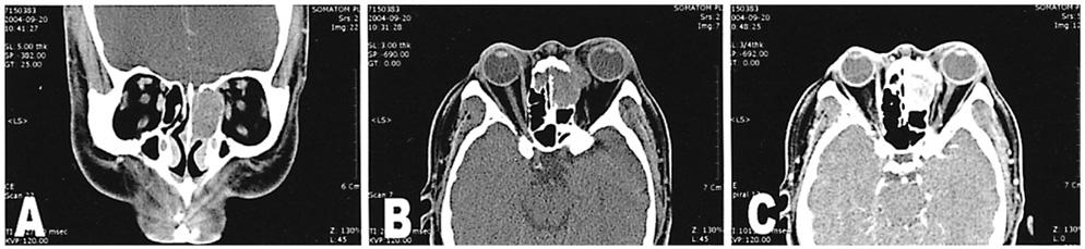
Fig. 1 Coronal (A) and axial (B,C) CT scans show a well-circumscribed, homogeneous mass in the left ethmoid sinus with extension into the left orbit and compression of the left medial rectus muscle. The tumor shows marked enhancement with contrast agents.
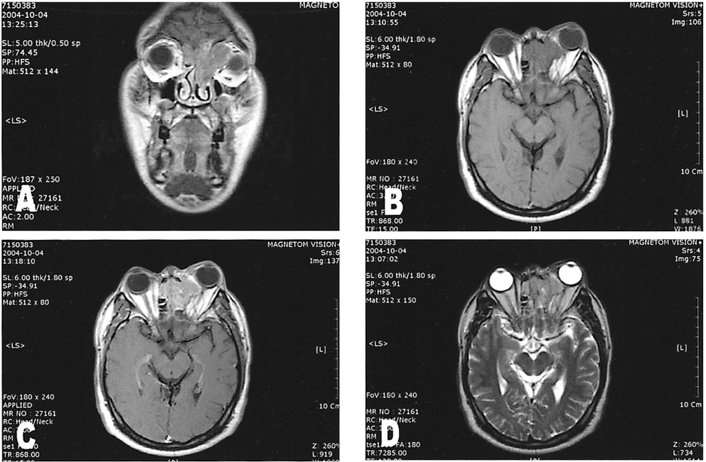
Fig. 2 Orbital magnetic resonance imaging (MRI) scans show rhabdomyosarcoma in left ethmoid sinus with low signal intensity on T1-weighted image (A,B) and moderate enhancement on enhanced scan (C). On a T2-weighted image, the lesion is isointense to extraocular muscles and hypointense to orbital fat (D). The lesion extends into the left orbit with compression of the left medial rectus muscle, and has intracranial extension with compression of the left side frontal lobe.
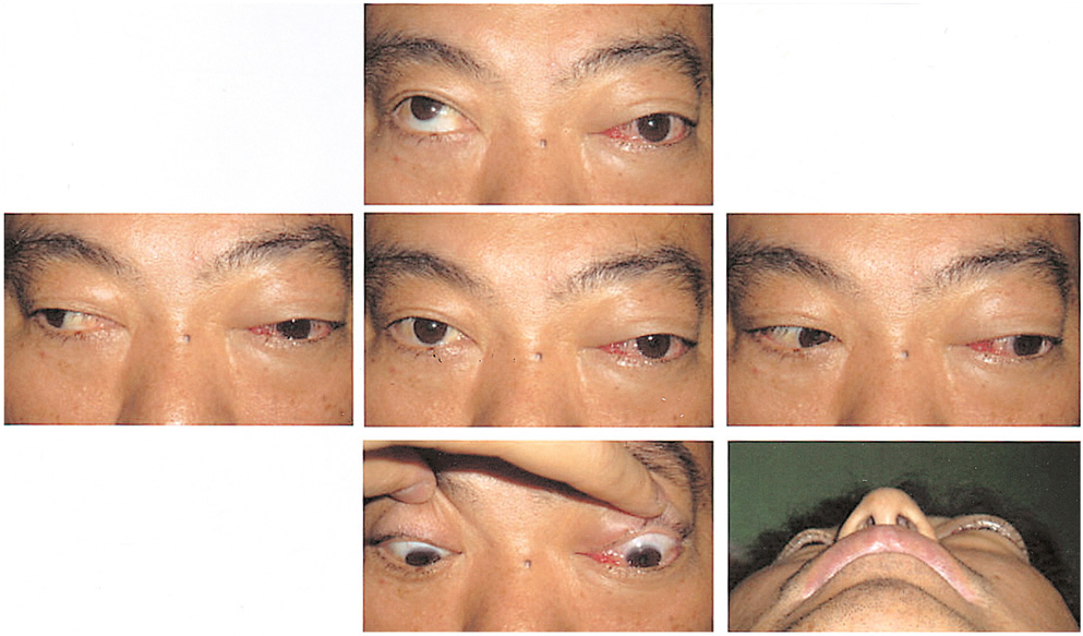
Fig. 3 Preoperative photography of a 48-year-old man suffering diplopia, with marked limitation of ocular movement, chemosis, proptosis in the left eye.
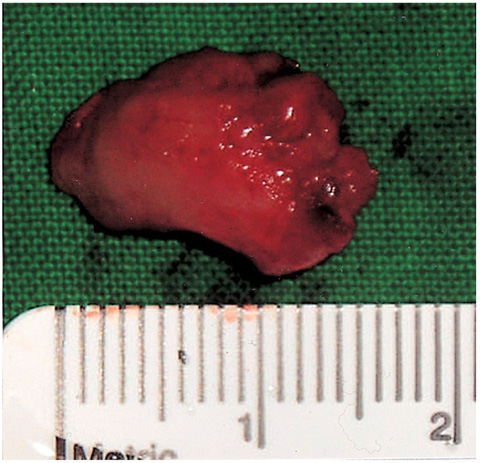
Fig. 4 Gross examination showed a circumscribed reddish tumor measuring 1.5×1.2×1.0 cm.

Fig. 5 Photomicrograph of isolated rhabdomyosarcoma. (A) Low magnification reveals diffuse small round cells (Hematoxylin-eosin, original magnification ×40). (B) High magnification reveals round cells (rhabdomyoblast) with eosinophilic cytoplasm and eccentric nuclei (arrow) (Hematoxylin-eosin, original magnification ×400). (C) Immunohistochemistry gave positive results with desmin, confirming the diagnosis of rhabdomyosarcoma (original magnification ×400). (D) Immunohistochemistry with smooth muscle actin (original magnification ×400).

Fig. 6 5-month postoperative photograph showing improvement of ocular movement and enophthalmos in the left eye.
Reference
-
1. Shields JA, Shields CL. Rhabdomyosarcoma: review for the ophthalmologist. Surv Ophthalmol. 2003. 48:39–57.2. Crist W, Gehan EA, Ragab AH, et al. The Third Intergroup Rhabdomyosarcoma Study. J Clin Oncol. 1995. 13:610–630.3. Lanzkowsky P. Rhabdomyosarcoma and other soft tissue sarcomas. Manual of Pediatric Hematology and Oncology. 2000. v. 1:3rd ed. New York: Academic Press;chap. 20.4. Karcioglu ZA, Hadjistilianou D, Rozans M, DeFrancesco S. Orbital rhabdomyosarcoma. Cancer Control. 2004. 11:328–333.5. Crist WM, Anderson JR, Meza JL, et al. Intergroup rhabdomyosarcoma study-IV: results for patients with nonmetastatic disease. J Clin Oncol. 2001. 19:3091–3102.6. Horn RC Jr, Enterline HT. Rhabdomyosarcoma: a clinicopathological study and classification of 39 cases. Cancer. 1958. 11:181–199.7. Little DJ, Ballo MT, Zagars GK, et al. Adult rhabdomyosarcoma: outcome following multimodality treatment. Cancer. 2002. 95:377–388.8. Esnaola NF, Rubin BP, Baldini EH, et al. Response to chemotherapy and predictors of survival in adult rhabdomyosarcoma. Ann Surg. 2001. 234:215–223.9. Hawkins WG, Hoos A, Antonescu CR, et al. Clinicopathologic analysis of patients with adult rhabdomyosarcoma. Cancer. 2001. 91:794–803.10. Friling R, Marcus M, Monos T, et al. Rhabdomyosarcoma: invading the orbit in an adult. Ophthal Plast Reconstr Surg. 1994. 10:283–286.11. Michael C. Stephen B, editor. Mesenchymal and fibro-osseous tumors. Principle and Practice of Ophthalmic Plastic and Reconstructive Surgery. 1996. v. 2. Philadelphia: W.B. Saunders;chap. 92.12. Shields CL, Shields JA, Honavar SG, Demirci H. Primary ophthalmic rhabdomyosarcoma in 33 patients. Trans Am Ophthalmol Soc. 2001. 99:133–142.13. Porterfield JF, Zimmerman LE. Rhabdomyosarcoma of the orbit; a clinicopathologic study of 55 cases. Virchows Arch Pathol Anat Physiol Klin Med. 1962. 335:329–344.14. Grieman RB, Katsikeris NF, Symington JM. Rhabdomyosarcoma of the maxillary sinus: review of the literature and report of a case. J Oral Maxillofac Surg. 1988. 46:1090–1096.15. Makishima K, Iwasaki H, Horie A. Alveolar rhabdomyosarcoma of the ethmoid sinus. Laryngoscope. 1975. 85:400–410.16. Sohaib SA, Moseley I, Wright JE. Orbital rhabdomyosarcoma: the radiological characteristics. Clin Radiol. 1998. 53:357–362.17. Koenigsberg RA, Noah R, Turtz A, et al. Rhabdomyosarcoma of the paranasal sinuses in an adult. Clin Imaging. 1995. 19:234–236.18. Shields CL, Shields JA, Honavar SG, Demirci H. Clinical spectrum of primary ophthalmic rhabdomyosarcoma. Ophthalmology. 2001. 108:2284–2292.
- Full Text Links
-
- Actions
-
Cited
- CITED
-
- Close
- Share
-
- Similar articles
-
- A Case of Ethmoid Sinus Mucocele with Transient Intraocular Pressure Elevation
- Traumatic Displacement of the Globe into the Ethmoid Sinus: Case Report
- A case of alveolar rhabdomyosarcoma of vulva
- Ectopic Meningioma of the Ethmoid Sinus: Report of a Case and Review of Literature
- A Case of Osteoma Involving the Orbit
