Korean J Ophthalmol.
2007 Sep;21(3):172-174. 10.3341/kjo.2007.21.3.172.
Bilateral Peripheral Infiltrative Keratitis After LASIK
- Affiliations
-
- 1Department of Ophthalmology, Kyung Hee University College of Medicine, Seoul, Korea. khjinmd@khmc.or.kr
- 2Department of Ophthalmology, KangWon University College of Medicine, KangWon do, Korea.
- 3Misolasik Ophthalmic Clinic, Suwon, Korea.
- KMID: 1099035
- DOI: http://doi.org/10.3341/kjo.2007.21.3.172
Abstract
- PURPOSE: To present a case of peripheral infiltrative keratitis mimicking infectious keratitis on the flap margin and limbus, which appeared on the first postoperative day after the laser in situ keratomileusis (LASIK). METHODS: A 36-year-old woman who underwent uneventful bilateral simultaneous LASIK developed multiple round infiltrate along the flap margin reaching to limbus from the 11 o'clock to 6 o'clock area in both eyes. RESULTS: The flap was lifted and irrigation was performed with antibiotics. but infiltration seemed to appear again. The infiltrate was more concentrated at the periphery and was extended to the limbus. Direct smear and culture for bacteria and fungus were negative. Topical prednisolone acetate 1% eye drops was added, infiltrative condition was resolved. CONCLUSIONS: LASIK induced peripheral infiltrative keratitis, in which infectious origin was ruled out, is reported.
Keyword
MeSH Terms
Figure
-
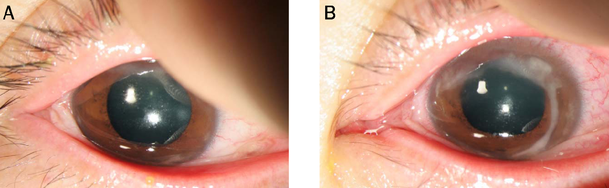
Fig. 1 White multiple corneal infiltrates in the periphery along the lamellar flap margin were noted in the first day after LASIK on both eyes (A: right eye, B: left eye)
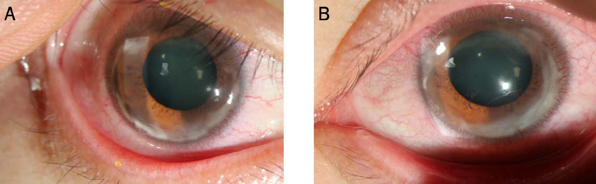
Fig. 2 Peripheral flap infiltration was progressed into the limbus in second day after flap lifting with antibiotics irrigation. (A: right eye, B: left eye)

Fig. 3 After adding topical prednisolone acetate along with topical antibiotics, decreased infiltration was the lamellar flap and the corneal limbus 6th day after flap lifting with antibiotics irrigation. (A: right eye, B: left eye)
Reference
-
1. Linebarger EJ, Hardten DR, Lindstrom RL. Diffuse lamellar keratitis : Diagnosis and management. J Cataract Refract Surg. 2000. 26:1072–1077.2. Linebarger EJ, Hardten DR, Lindstrom RL. Diffuse lamellar keratitis: identification and management. Int Ophthalmol Clin. 2000. 40:77–86.3. Blustein JN, Hitchins VM, Woo EK. Diffuse lamellar keratitis, endotoxin, and ophthalmic sponges. J Cataract Refract Surg. 2004. 30:2027–2028.4. Lifshitz T, Levy J, Mahler O, Levinger S. Peripheral sterile corneal infiltrates after refractive surgery. J Cataract Refract Surg. 2005. 31:1392–1395.5. Lahner WJ, Hardten DR, Lindstrom RL. Peripheral keratitis following laser in situ keratomileusis. J Refract Surg. 2003. 19:671–675.6. Singhal S, Sridhar MS, Garg P. Bilateral peripheral infiltrative keratitis after LASIK. J Refract Surg. 2005. 21:402–404.7. Ambrosio R, Periman LM, Netto MV, Wilson SE. Bilateral marginal sterile infiltrates and diffuse lamellar keratitis after laser in situ keratomileusis. J Refract Surg. 2003. 19:154–158.8. Holzer MP, Solomon KD, Vroman DT, et al. Diffuse lamellar keratitis: evaluation of etiology, histopathologic findings, and clinical implications in an experimental animal model. J Cataract Refract Surg. 2003. 29:542–549.
- Full Text Links
-
- Actions
-
Cited
- CITED
-
- Close
- Share
-
- Similar articles
-
- A Case of Sterile Peripheral Corneal Infiltrative Event after LASIK
- Suspected Fungal Keratitis After LASIK: Treated with Flap Removal and Medical Therapy
- Clinical Analysis of Infectious Keratitis after LASIK
- A Case of Mycobacterium Fortuitum Keratitis at the Interface of the Cornea after LASIK
- A Case of Congenital Syphilitic Interstitial Keratitis
