A Simple New Method for Identifying the Proximal Cut End in Lower Canalicular Laceration
- Affiliations
-
- 1Department of Ophthalmology, College of Medicine, Konyang University, Daejeon, Korea. hmseye@hanmail.net
- KMID: 1098998
- DOI: http://doi.org/10.3341/kjo.2008.22.2.73
Abstract
- PURPOSE: We report a simple and effective method of identifying the medial cut end of lower canalicular laceration cases. METHODS: Twenty-seven eyes with lower canalicular lacerations as a result of trauma were involved in the study. Surgery was performed within 48 hours after injury for canalicular reconstruction. Upper canalicular probing was utilized to identify the medial cut end of deep canalicular lacerations when difficulties were encountered. Total time from the initiation of the probing procedure to the identification of the medial cut end of the lower canaliculus was measured. RESULTS: A total of 27 eyes with lower canalicular lacerations were reconstructed. In 20 eyes, the medial lacerated end was located by upper canalicular probing. The mean time from initiation of the probing procedure to identification of the medial cut end of the lacerated canaliculus was 2 minutes. CONCLUSIONS: We conclude that upper canalicular probing in patients with lower canalicular lacerations significantly reduces the time from the initiation of the operation to the identification of the medial cut end of the lower canaliculus.
MeSH Terms
Figure
-

Fig. 1 (A) The lacrimal probe is exposed through the medial cut end of the lacerated canaliculi. (B) The common canaliculus is pushed towards the operative field by the lacrimal probe. (C) Postoperative state 1 month after operation. The silicone tube is visible between the upper and lower canaliculi.
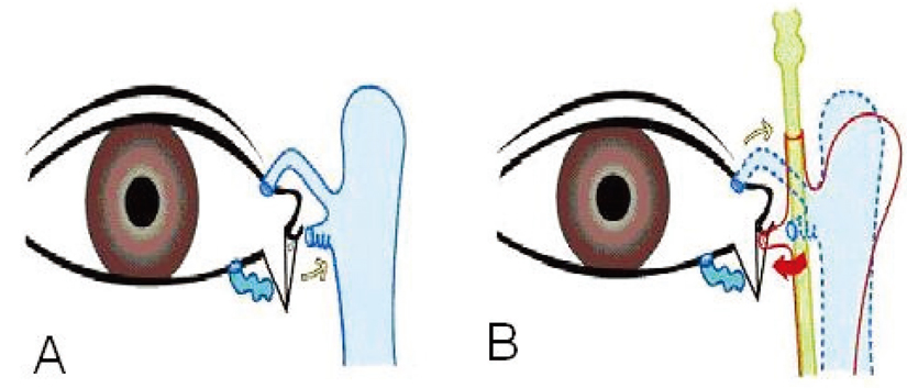
Fig. 2 (A) The medial cut end of the lacerated lower canaliculus is retracted towards the lacrimal sac (yellow arrow) after injury. (B) By upper punctal probing (yellow arrow), the medial cut end is pushed towards the lacerated skin margin (red arrow).
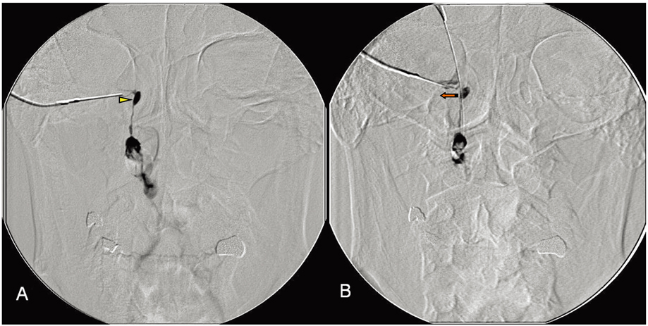
Fig. 3 DCG demonstrates that the lacrimal sac and canaliculus are lateralized after probing. The lateral border of the lacrimal sac (yellow arrow head) before lacrimal probing (A) is moved laterally (red arrow) after lacrimal probing (B).
Reference
-
1. David M, Reifler DM. Management of canalicular laceration. Surv Ophthalmol. 1991. 36:113–132.2. Worst JG. Method for reconstructing torn lacrimal canaliculus. Am J Ophthalmol. 1962. 53:520–522.3. Hing SJ. A retrospective study of lacrimal canaliculaus injuries in Auckland. Trans Ophthalmol Soc N Z. 1984. 36:72–73.4. Jordan DR, Nerad JA, Tse DT. The pigtail probe, revisited. Ophthalmology. 1990. 97:512–519.5. Saunders DH, Shannon GM, Flanagan JC. The effectiveness of the pigtail probe method of repairing canalicular lacerations. Ophthalmic Surg. 1978. 9:33–40.6. Welham RAN. The immediate management of injuries to the lacrimal drainage apparatus. Trans Ophthalmol Soc U K. 1982. 102:216–217.7. Morrison FD. An aid to repair of lacerated tear ducts. Arch Ophthalmol. 1964. 72:341–342.8. Loff HJ, Wobig JL, Dailey RA. The bubble test: an atraumatic method for canalicular laceration repair. Ophthal Plast Reconstr Surg. 1996. 12(1):61–64.9. Hawes MJ, Dortzbach RK. lindberg JV, editor. Trauma of the lacrimal drainage system. Lacrimal surgery. 1988. New York: Churchill Livingstone;241–262.10. Kersten RC, Kulwin DR. "One stich" Canalicular repair. A simplified approach for repair of canalicular laceration. Ophthalmology. 1996. 103:785–789.
- Full Text Links
-
- Actions
-
Cited
- CITED
-
- Close
- Share
-
- Similar articles
-
- Delayed Lacrimal Stent Implantation Using Mini Monoka(R) in Canalicular Laceration
- Clinical Experience of Monocanaliculonasal Stent in Canalicular Laceration
- Bicanalicular Silicone Intubation of Canalicular Laceration
- Clinical Experience of Canaliculaoplasty Using Mini-Monoka
- Clinical Features Associated With Outcomes of Canalicular Laceration Repair
