Physeal Growth Arrest by Excessive Compression: Histological, Biochemical, and Micro-CT Observations in Rabbits
- Affiliations
-
- 1Department of Orthopedic Surgery, Seoul National University Children's Hospital, Seoul National University College of Medicine, Seoul, Korea. inhoc@snu.ac.kr
- 2Department of Radiology, Seoul National University Children's Hospital, Seoul National University College of Medicine, Seoul, Korea.
- KMID: 1097169
- DOI: http://doi.org/10.4055/cios.2011.3.4.309
Abstract
- BACKGROUND
Compressive force across the growth plate may cause retardation and even arrest of physeal growth. The purpose of this study was to investigate histologic changes, metabolic changes in terms of glycosaminoglycan (GAG) concentration, and contrast-enhanced micro-computed tomography (CEMCT) findings of physeal cartilage in a rabbit model of physeal damage caused by excessive compression.
METHODS
Compressive forces were applied via external fixators for two weeks to the growth plates of distal femurs and proximal tibiae of right hind-legs in 8-week-old rabbits. Left hind-legs remained intact and were used as controls. Forty-four bone specimens containing growth plates of distal femurs or proximal tibiae were harvested one week (n = 12) and four weeks (n = 32) after surgery, and examined for histologic findings (H&E staining) and GAGs quantification in physeal cartilage. After incubation in an ionic contrast material for 48 hours, specimens were scanned by CEMCT, and the pixel values of physeal cartilage were measured.
RESULTS
CEMCT showed a thin, highly attenuated line parallel to the growth plate in compressed specimens harvested at four weeks after surgery, which was found to be transversely connected trabecular bone. In these specimens, GAG content in physeal cartilage was significantly lower, and CEMCT pixel values of physeal cartilage were significantly higher than in the specimens from the contralateral control side.
CONCLUSIONS
Excessive compressive force applied to growth plates produces altered histologic features and metabolic function in terms of decreased GAG content in physeal cartilage, changes that can be demonstrated by CEMCT.
MeSH Terms
Figure
-
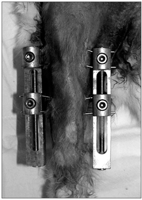
Fig. 1 Two custom-made external fixators were used to apply compressive force to the growth plate of the distal femur and proximal tibia across the knee joint in the right hind-leg.
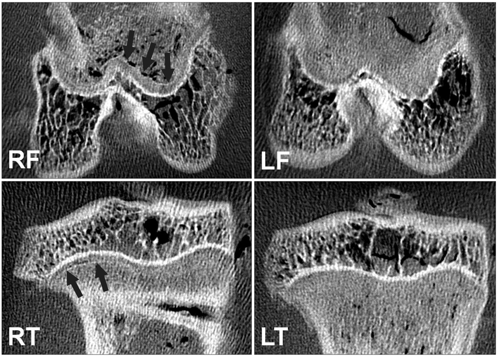
Fig. 2 Contrast-enhanced micro-computed tomography images showed that a thin, highly attenuated line parallel to the growth plate (arrows) was formed in compressed specimens (RF and RT) harvested at four weeks after surgery. RF: right femur, RT: right tibia, LF: left femur, LT: left tibia.

Fig. 3 A histologic section of a compressed specimen harvested at four weeks after surgery showed newly formed trabecular bones that were interconnected haphazardly (asterisk) and transversely connected trabecular bone (arrows), the latter of which corresponded with the highly attenuated line observed in the contrast-enhanced micro-computed tomography (A: × 10, B: H&E, × 100).
Reference
-
1. Futami T, Foster BK, Morris LL, LeQuesne GW. Magnetic resonance imaging of growth plate injuries: the efficacy and indications for surgical procedures. Arch Orthop Trauma Surg. 2000. 120(7-8):390–396.
Article2. Cockman MD, Blanton CA, Chmielewski PA, et al. Quantitative imaging of proteoglycan in cartilage using a gadolinium probe and microCT. Osteoarthritis Cartilage. 2006. 14(3):210–214.
Article3. Palmer AW, Guldberg RE, Levenston ME. Analysis of cartilage matrix fixed charge density and three-dimensional morphology via contrast-enhanced microcomputed tomography. Proc Natl Acad Sci U S A. 2006. 103(51):19255–19260.
Article4. Piscaer TM, van Osch GJ, Verhaar JA, Weinans H. Imaging of experimental osteoarthritis in small animal models. Biorheology. 2008. 45(3-4):355–364.
Article5. Farndale RW, Buttle DJ, Barrett AJ. Improved quantitation and discrimination of sulphated glycosaminoglycans by use of dimethylmethylene blue. Biochim Biophys Acta. 1986. 883(2):173–177.
Article6. Harris HA. Bone growth in health and disease. 1933. London: Oxford University Press.7. Ogden JA. The evaluation and treatment of partial physeal arrest. J Bone Joint Surg Am. 1987. 69(8):1297–1302.
Article8. Shapiro F. Pediatric orthopedic deformities: basic science, diagnosis, and treatment. 2001. San Diego: Academic Press;555.9. Moyer HR, Wang Y, Farooque T, et al. A new animal model for assessing cartilage repair and regeneration at a nonarticular site. Tissue Eng Part A. 2010. 16(7):2321–2330.
Article10. Knudson CB, Knudson W. Cartilage proteoglycans. Semin Cell Dev Biol. 2001. 12(2):69–78.
Article11. Roughley PJ. The structure and function of cartilage proteoglycans. Eur Cell Mater. 2006. 12:92–101.
Article12. Cancel M, Grimard G, Thuillard-Crisinel D, Moldovan F, Villemure I. Effects of in vivo static compressive loading on aggrecan and type II and X collagens in the rat growth plate extracellular matrix. Bone. 2009. 44(2):306–315.
Article13. Bonnel F, Peruchon E, Baldet P, Dimeglio A, Rabischong P. Effects of compression on growth plates in the rabbit. Acta Orthop Scand. 1983. 54(5):730–733.
Article14. Langenskiold A. Traumatic premature closure of the distal tibial epiphyseal plate. Acta Orthop Scand. 1967. 38(4):520–531.
Article15. Hasler CC, Foster BK. Secondary tethers after physeal bar resection: a common source of failure? Clin Orthop Relat Res. 2002. (405):242–249.16. Foster BK, John B, Hasler C. Free fat interpositional graft in acute physeal injuries: the anticipatory Langenskiold procedure. J Pediatr Orthop. 2000. 20(3):282–285.
Article
- Full Text Links
-
- Actions
-
Cited
- CITED
-
- Close
- Share
-
- Similar articles
-
- Experimental Study for the Effects of Free Physeal Transplantation to Regain Bone Grwoth after Partial Physeal Injury
- Physeal Growth Arrest Caused by Thromboembolism of the Right Femoral Artery in a Premature Infant
- Salter-Harris Type IV Physeal Fracture of the Distal Radius: A Case Report
- Cultured Chondrocyte Transplantation in the Damaged Growth Plate
- Prognostic Factors of Physeal Bar Resection and Fat Graft Interposition in the Treatment of Partial Physeal Arrest
