Inflammatory Pseudotumor of the Breast: a Case Report with Imaging Findings
- Affiliations
-
- 1Seoul National University Hospital Healthcare System Gangnam Center, Seoul 135-984, Korea.
- 2Department of Radiology and Clinical Research Institute, Seoul National University Hospital and the Institute of Radiation Medicine, Seoul National University Medical Research Center, Seoul 110-744, Korea. moonwk@radcom.snu.ac.kr
- 3Department of Pathology, University of Ulsan College of Medicine, Asan Medical Center, Seoul 138-736, Korea.
- KMID: 1093968
- DOI: http://doi.org/10.3348/kjr.2009.10.5.515
Abstract
- Inflammatory pseudotumor, also known as inflammatory myofibroblastic tumor and plasma cell granuloma, is an uncommon low-grade lesion composed of spindle cells admixed with mature plasma cells and other inflammatory cells, such as histiocytes, lymphocytes, and eosinophils. Here, we describe the mammographic and ultrasonographic findings of a case of an inflammatory pseudotumor of the breast in a 60-year-old woman. With the suspicion of malignancy, core needle biopsy and surgical excision confirmed the mass as being an inflammatory pseudotumor of the breast.
Keyword
MeSH Terms
Figure
-
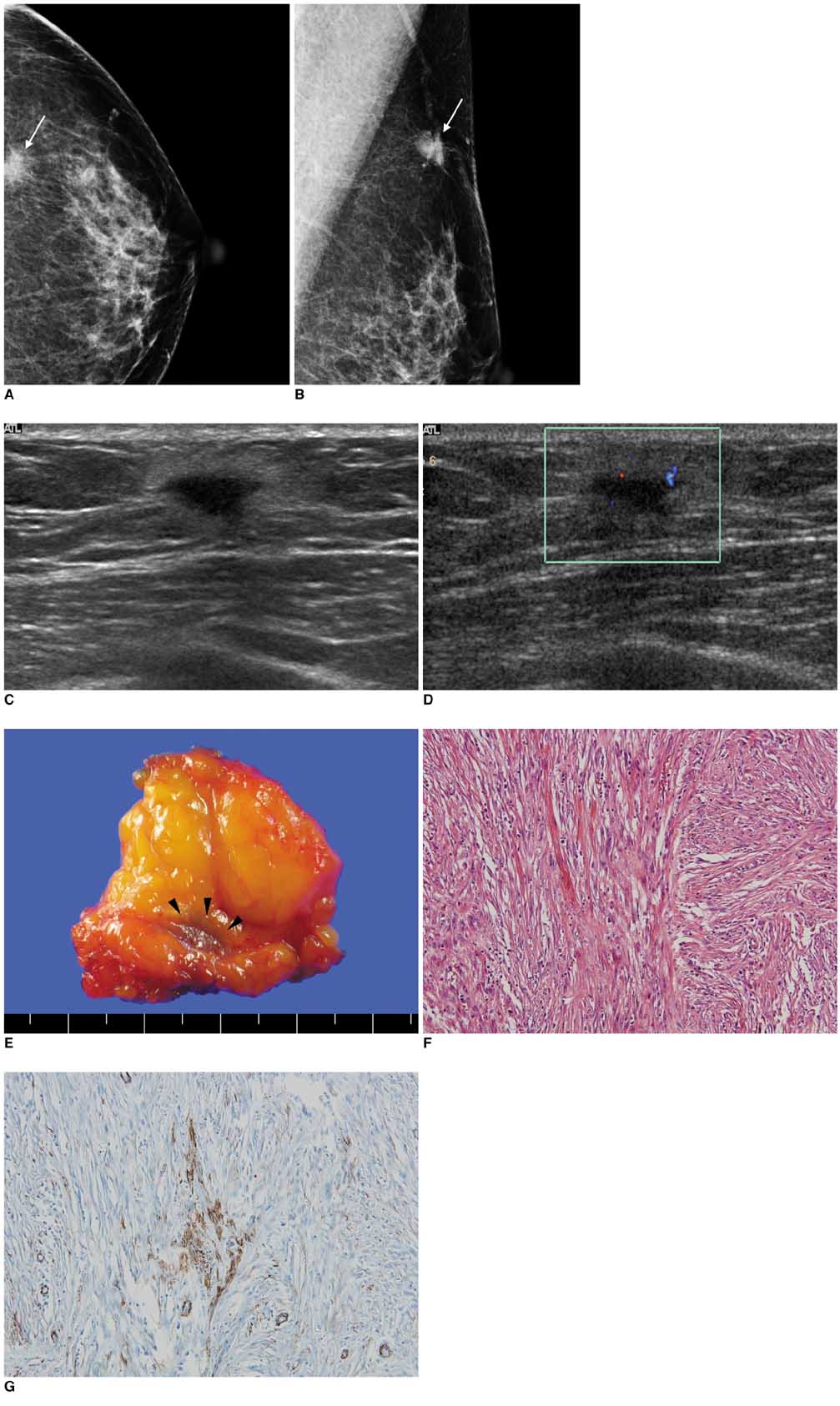
Fig. 1 Inflammatory pseudotumor of breast in 60-year-old woman. A, B. Left craniocaudal (A) and mediolateral oblique (B) mammograms reveal 1.5-cm-sized ill-defined, high-density mass (arrows) in axillary tail area of left breast. C. Transverse US scan reveals irregular shaped, ill-defined homogeneous hypoechoic mass with echogenic halo in left axillary tail region. We found nodule surrounded by fat lobules and mass appearing to infiltrate around fat lobules. D. Color Doppler study reveals moderate vascularity in peripheral halo portion of mass. E. Upon gross pathology, we observed ill-defined pinkish-white mass (arrowheads) without necrosis or hemorrhage. F. For microscopic findings at high magnification, proliferating spindle cells had bland-looking nuclei and nucleoli were inconspicuous. There were occasional mitoses (up to 3 of 10 per high-power field), but atypical mitoses were not found (Hematoxylin & Eosin staining, ×200). G. Following immunohistochemical assay, spindle cells were found to be reactive for anti-SMA (smooth muscle actin), which demonstrates myofibroblastic differentiation (×200).
Reference
-
1. Haj M, Weiss M, Loberant N, Cohen I. Inflammatory pseudotumor of the breast: case report and literature review. Breast J. 2003. 9:423–425.2. Yip CH, Wong KT, Samuel D. Bilateral plasma cell granuloma (inflammatory pseudotumour) of the breast. Aust N Z J Surg. 1997. 67:300–302.3. Zardawi IM, Clark D, Williamsz G. Inflammatory myofibroblastic tumor of the breast. A case report. Acta Cytol. 2003. 47:1077–1081.4. Khanafshar E, Phillipson J, Schammel DP, Minobe L, Cymerman J, Weidner N. Inflammatory myofibroblastic tumor of the breast. Ann Diagn Pathol. 2005. 9:123–129.5. Ilvan S, Celik V, Paksoy M, Cetinaslan I, Calay Z. Inflammatory myofibroblastic tumor (inflammatory pseudotumor) of the breast. APMIS. 2005. 113:66–69.6. Zen Y, Kasahara Y, Horita K, Miyayama S, Miura S, Kitagawa S, et al. Inflammatory pseudotumor of the breast in a patient with a high serum IgG4 level: histologic similarity to sclerosing pancreatitis. Am J Surg Pathol. 2005. 29:275–278.7. Sastre-Garau X, Couturier J, Derre J, Aurias A, Klijanienko J, Lagace R. Inflammatory myofibroblastic tumour (inflammatory pseudotumour) of the breast. Clinicopathological and genetic analysis of a case with evidence for clonality. J Pathol. 2002. 196:97–102.8. Chetty R, Govender D. Inflammatory pseudotumor of the breast. Pathology. 1997. 29:270–271.9. Pettinato G, Manivel JC, Insabato L, De Chiara A, Petrella G. Plasma cell granuloma (inflammatory pseudotumor) of the breast. Am J Clin Pathol. 1988. 90:627–632.10. Coffin CM, Watterson J, Priest JR, Dehner LP. Extrapulmonary inflammatory myofibroblastic tumor (inflammatory pseudotumor). A clinicopathologic and immunohistochemical study of 84 cases. Am J Surg Pathol. 1995. 19:859–872.11. Pettinato G, Manivel JC, De Rosa N, Dehner LP. Inflammatory myofibroblastic tumor (plasma cell granuloma). Clinicopathologic study of 20 cases with immunohistochemical and ultrastructural observations. Am J Clin Pathol. 1990. 94:538–546.12. Maier HC, Sommers SC. Recurrent and metastatic pulmonary fibrous histiocytoma/plasma cell granuloma in a child. Cancer. 1987. 60:1073–1076.13. Cessna MH, Zhou H, Sanger WG, Perkins SL, Tripp S, Pickering D, et al. Expression of ALK1 and p80 in inflammatory myofibroblastic tumor and its mesenchymal mimics: a study of 135 cases. Mod Pathol. 2002. 15:931–938.14. Freeman A, Geddes N, Munson P, Joseph J, Ramani P, Sandison A, et al. Anaplastic lymphoma kinase (ALK1) staining and molecular analysis in inflammatory myofibroblastic tumours of the bladder: a preliminary clinicopathological study of nine cases and review of the literature. Mod Pathol. 2004. 17:765–771.15. Akbulut M, Gunhan-Bilgen I, Zekioglu O, Duygulu G, Oktay A, Ozdemir N. Fine needle aspiration cytology of inflammatory myofibroblastic tumour (inflammatory pseudotumour) of the breast: a case report and review of the literature. Cytopathology. 2007. 18:384–387.
- Full Text Links
-
- Actions
-
Cited
- CITED
-
- Close
- Share
-
- Similar articles
-
- A Case of Inflammatory Pseudotumor Cerebri and Nasal Septum
- A Case of Inflammatory Pseudotumor in the Retroperitoneum
- Inflammatory Myofibroblastic Tumor of the Breast: A Case Report
- Inflammatory Pseudotumor in the Liver and Right Omentum Caused by Pelvic Inflammatory Disease: A Case Report
- Inflammatory Pseud0tumor of the Liver: A case report
