Sonographic Findings of Uterine Endometrial Stromal Sarcoma
- Affiliations
-
- 1Department of Radiology, Cheil General Hospital, Sungkyunkwan University School of Medicine, Seoul, Korea. chrismd@hanmail.net
- 2Department of Diagnostic Pathology, Cheil General Hospital, Sungkyunkwan University School of Medicine, Seoul, Korea.
- KMID: 1092552
- DOI: http://doi.org/10.3348/kjr.2006.7.4.281
Abstract
OBJECTIVE
The study was performed to present the sonographic findings of uterine endometrial stromal sarcoma (ESS). MATERIALS AND METHODS: We conducted a retrospective review of sonographic findings of 10 cases that were diagnosed as uterine ESS. The patients' ages ranged from 25 to 51 years (mean age: 36.1 years). The reviews focused on the location, margin, size, number and echotexture of the lesions. Hysterectomy (n = 9) and myomectomy (n = 1) were performed and a pathologic diagnosis was obtained in all cases. RESULTS: The masses were located in the uterine wall (n = 6), or they presented as a polypoid mass protruding into the endometrial cavity from the myometrium (n = 3) or as a central cavity mass (n = 1). The lesion margins were smooth (n = 5), ill defined (n = 2), or smooth with partially nodular extensions (n = 3). The maximal mass length was 38 mm to 160 mm with a mean mass length of 83.5 mm. There were single lesions in eight cases and multiple lesions in two cases. The lesion echotextures were hypoechoic solid (n = 3), heterogeneously intermediate echoic (n = 5), diffuse myometrial thickening with heterogeneous echogenicity (n = 1) and septated cystic (n = 1). CONCLUSION: Endometrial stromal sarcoma presents with four patterns of its sonographic appearance; a polypoid mass with nodular myometrial extension, an intramural mass with an ill defined margin and heterogeneous echogenicity, an ill defined large central cavity mass or, diffuse myometrial thickening.
MeSH Terms
Figure
-
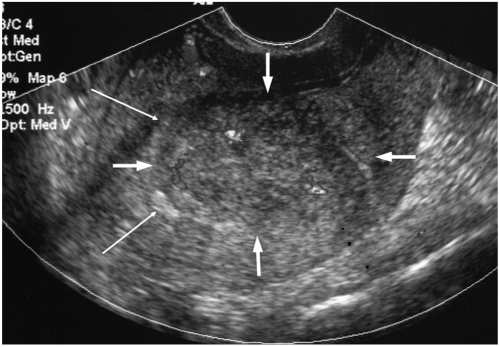
Fig. 1 Sonographic findings of endometrial stromal sarcoma that presented as a mural mass in patient 2.A heterogeneously echoic mass with an ill defined margin is noted in the uterine wall (thick arrows). The endometrium is displaced by the mass (thin arrows). Color Doppler sonography demonstrates a focally dispersed vascularity within the mass.Pathology revealed a subendometrial endometrial stromal sarcoma invading more than half of the myometrial thickness (not shown).
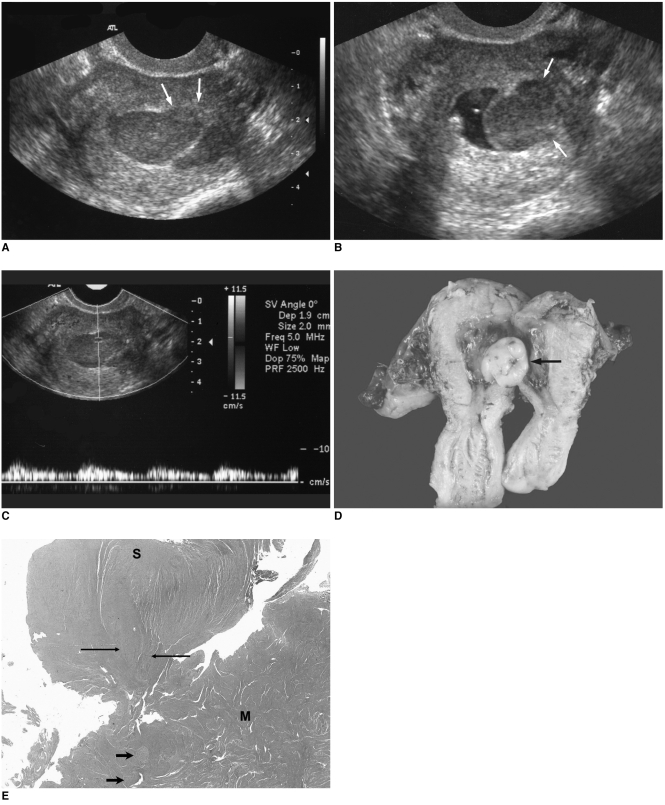
Fig. 2 Sonographic findings of endometrial stromal sarcoma, which presented as a polypoid mass protruding into the endometrial cavity from the myometrium in patient 1.
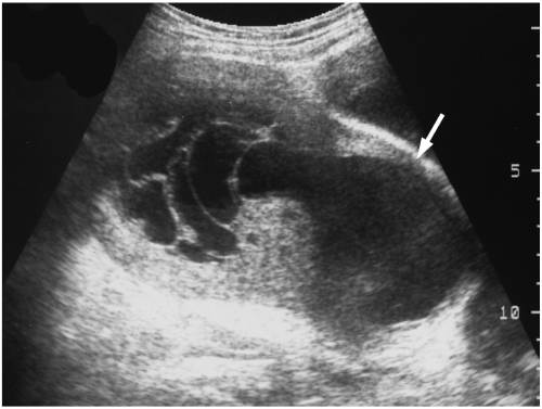
Fig. 3 Endometrial stromal sarcoma that presented as a huge central cavity mass.An ill defined, septated cystic mass with a solid portion is noted in the uterine central cavity in patient 9. The endometrium is obliterated by the mass and myometrial thinning is noted (arrow).Pathology revealed a low grade endometrial stromal sarcoma invading the endometrium and almost the full thickness of the anterior myometrium and cystic degeneration of the tumor (not shown).
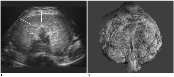
Fig. 4 Endometrial stromal sarcoma that presented with diffuse myometrial thickening.
Reference
-
1. Kahanpaa KV, Wahlstrom T, Grohn P, Heinonen E, Nieminen U, Widholm O. Sarcoma of the uterus: a clinicopathologic study of 119 patients. Obstet Gynecol. 1989; 67:417–424. PMID: 3945454.2. Evans HL. Endometrial stromal sarcoma and poorly differentiated endometrial sarcoma. Cancer. 1982; 50:2170–2182. PMID: 7127257.
Article3. Chen CD, Huang CC, Wu CC, Tseng GC, Lee CN, Lin GJ, et al. Sonographic characteristics in low-grade endometrial stromal sarcoma: a report of two cases. J Ultrasound Med. 1995; 14:165–168. PMID: 8568965.
Article4. Tepper R, Altaras M, Goldberger S, Zalel Y, Cordoba M, Beyth Y. Color Doppler ultrasonographic findings in low and high grade endometrial stromal sarcomas. J Ultrasound Med. 1994; 13:817–819. PMID: 7823348.
Article5. Carter J, Perrone T, Carson LF, Carlson J, Twiggs LB. Uterine malignancy predicted by transvaginal sonography and color flow Doppler ultrasonography. J Clin Ultrasound. 1993; 21:405–408. PMID: 8227385.
Article6. Kaminski PF, Podczaski ES. Hernandez E, Atkinson BF, editors. Premalignant and malignant conditions of the uterus. Clinical gynecologic pathology. 1996. Philadelphia, Pennsylvania: W.B. Saunders;p. 334–339.7. Oliva E, Clement PB, Young RH. Endometrial stromal tumors: an update on a group of tumors with a protean phenotype. Adv Anat Pathol. 2000; 7:257–281. PMID: 10976906.
Article8. Norris HJ, Taylor HB. Mesenchymal tumors of the uterus. I: A clinical and pathological study of 53 endometrial stromal tumors. Cancer. 1966; 19:755–766. PMID: 5939046.
Article9. Koyama T, Togashi K, Konishi I, Kobayashi H, Itoh T, Higuchi T, et al. MR imaging of endometrial stromal sarcoma: correlation with pathologic findings. AJR Am J Roentgenol. 1999; 173:767–772. PMID: 10470920.
Article10. Ueda M, Otsuka M, Hatakenaka M, Sakai S, Ono M, Yoshimitsu K, et al. MR imaging findings of uterine endometrial stromal sarcoma: differentiation from endometrial carcinoma. Eur Radiol. 2001; 11:28–33. PMID: 11194912.
Article11. Gandolfo N, Gandolfo NG, Serafini G, Martinoli C. Endometrial stromal sarcoma of the uterus: MR and US findings. Eur Radiol. 2000; 10:776–779. PMID: 10823632.
Article12. Perez-Montiel D, Salmeron AA, Malagon HD. Multicystic endometrial stromal sarcoma. Ann Diagn Pathol. 2004; 8:213–218. PMID: 15290672.13. Cacciatore B, Lehtovirta P, Wahlstrom T, Ylostalo P. Ultrasound findings in uterine mixed Mullerian sarcomas and endometrial stromal sarcomas. Gynecol Oncol. 1989; 35:290–293. PMID: 2689303.
Article14. Callen PW. Richenberg J, Cooperberg P, editors. Ultrasound of the uterus. Ultrasonography in obstetrics and gynecology. 2000. 4th ed. Philadelphia, Pennsylvania: W.B. Saunders;p. 837–838.
- Full Text Links
-
- Actions
-
Cited
- CITED
-
- Close
- Share
-
- Similar articles
-
- A Case of Low-Grade Endometrial Stromal Sarcoma of the Uterus (So-Called ""Endolymphatic Stromal Myosis"")
- Two Cases of Low Grade Endometrial Stromal Sarcoma
- Two Cases of Low-Grade Endometrial Stromal Sarcoma
- A case of primary retroperitoneal undifferentiated endometrial stromal sarcoma after concurrent chemoradiation therapy for cervical cancer
- A Case of Post-total hysterectomy with bilat. salpingooophorectomy Retroperitoneal Endometrial Stromal Sarcoma
