Korean J Radiol.
2006 Dec;7(4):243-248. 10.3348/kjr.2006.7.4.243.
Volumetric Measurements of Lung Nodules with Multi-Detector Row CT: Effect of Changes in Lung Volume
- Affiliations
-
- 1Department of Radiology, Seoul National University College of Medicine, and the Institute of Radiation Medicine, SNUMRC, Seoul, Korea. jmgoo@plaza.snu.ac.kr
- 2Mallinckrodt Institute of Radiology, Washington University School of Medicine, 510 S Kingshighway Blvd, St Louis, MO 63110, USA.
- 3Division of Pulmonary and Critical Care Medicine, Department of Internal Medicine, Washington University School of Medicine, 510 S Kingshighway Blvd, St Louis, MO 63110, USA.
- KMID: 1092547
- DOI: http://doi.org/10.3348/kjr.2006.7.4.243
Abstract
OBJECTIVE
To evaluate how changes in lung volume affect volumetric measurements of lung nodules using a multi-detector row CT. MATERIALS AND METHODS: Ten subjects with asthma or chronic bronchitis who had one or more lung nodules were included. For each subject, two sets of CT images were obtained at inspiration and at expiration. A total of 33 nodules (23 nodules > or = 3 mm) were identified and their volume measured using a semiautomatic volume measurement program. Differences between nodule volume on inspiration and expiration were compared using the paired t-test. Percent differences, between on inspiration and expiration, in nodule attenuation, total lung volume, whole lung attenuation, and regional lung attenuation, were computed and compared with percent difference in nodule volume determined by linear correlation analysis. RESULTS: The difference in nodule volume observed between inspiration and expiration was significant (p < 0.01); the mean percent difference in lung nodule volume was 23.1% for all nodules and for nodules > or = 3 mm. The volume of nodules was measured to be larger on expiration CT than on inspiration CT (28 out of 33 nodules; 19 out of 23 nodules > or = 3 mm). A statistically significant correlation was found between the percent difference of lung nodule volume and lung volume or regional lung attenuation (p < 0.05) for nodules > or = 3 mm. CONCLUSION: Volumetric measurements of pulmonary nodules were significantly affected by changes in lung volume. The variability in this respiration-related measurement should be considered to determine whether growth has occurred in a lung nodule.
Keyword
MeSH Terms
Figure
-
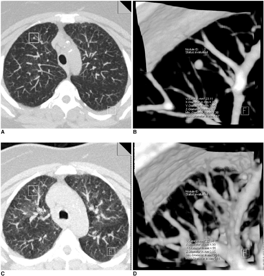
Fig. 1 Maximal intensity projection (A, C) and volume rendering (B, D) images on inspiration (A, B) and expiration (C, D). Percent differences for the lung nodule volume, nodule attenuation, lung volume, whole lung attenuation, and regional lung attenuation are 34.8%, 57.8%, 73.1%, 17.3%, and 20.5%, respectively.
Reference
-
1. Fischbach F, Knollmann F, Griesshaber V, Freund T, Akkol E, Felix R. Detection of pulmonary nodules by multislice computed tomography: improved detection rate with reduced slice thickness. Eur Radiol. 2003. 13:2378–2383.2. Benjamin MS, Drucker EA, McLoud TC, Shepard JA. Small pulmonary nodules: detection at chest CT and outcome. Radiology. 2003. 226:489–493.3. Goo JM, Chung MJ, Lee HJ, Im JG. Posterior subpleural nodules in patients with underlying malignancies: value of prone computed tomography. J Comput Assist Tomogr. 2003. 27:274–278.4. Swensen SJ, Jett JR, Hartman TE, Midthun DE, Sloan JA, Sykes AM, et al. Lung cancer screening with CT: Mayo Clinic experience. Radiology. 2003. 226:756–761.5. Diederich S, Thomas M, Semik M, Lenzen H, Roos N, Weber A, et al. Screening for early lung cancer with low-dose spiral computed tomography: results of annual follow-up examinations in asymptomatic smokers. Eur Radiol. 2004. 14:691–702.6. Yankelevitz DF, Reeves AP, Kostis WJ, Zhao B, Henschke CI. Small pulmonary nodules: volumetrically determined growth rates based on CT evaluation. Radiology. 2000. 217:251–256.7. Ko JP, Rusinek H, Jacobs EL, Babb JS, Betke M, McGuinness G, et al. Small pulmonary nodules: volume measurement at chest CT-phantom study. Radiology. 2003. 228:864–887.8. Winer-Muram HT, Jennings SG, Meyer CA, Liang Y, Aisen AM, Tarver RD, et al. Effect of varying CT section width on volumetric measurement of lung tumors and application of compensatory equations. Radiology. 2003. 229:184–194.9. Goo JM, Tongdee T, Tongdee R, Yeo K, Hildebolt CF, Bae KT. Volumetric measurement of synthetic lung nodules with multidetector row CT: effect of various image reconstruction parameters and segmentation thresholds on measurement accuracy. Radiology. 2005. 235:850–856.10. Revel MP, Lefort C, Bissery A, Bienvenu M, Aycard L, Chatellier G, et al. Pulmonary nodules: preliminary experience with three-dimensional evaluation. Radiology. 2004. 231:459–466.11. Wormanns D, Kohl G, Klotz E, Marheine A, Beyer F, Heindel W, et al. Volumetric measurements of pulmonary nodules at multi-row detector CT: in vivo reproducibility. Eur Radiol. 2004. 14:86–92.12. Standards for the diagnosis and care of patients with chronic obstructive pulmonary disease (COPD) and asthma. This official statement of the American Thoracic Society was adopted by the ATS Board of Directors, November 1986. Am Rev Respir Dis. 1987. 136:225–244.13. Proceedings of the ATS workshop on refractory asthma: current understanding, recommendations, and unanswered questions. American Thoracic Society. Am J Respir Crit Care Med. 2000. 162:2341–2351.14. Gierada DS, Yusen RD, Pilgram TK, Crouch L, Slone RM, Bae KT, et al. Repeatability of quantitative CT indexes of emphysema in patients evaluated for lung volume reduction surgery. Radiology. 2001. 220:448–454.15. Goo JM, Lee JW, Lee HJ, Kim S, Kim JH, Im JG. Automated lung nodule detection at low-dose CT: preliminary experience. Korean J Radiol. 2003. 4:211–216.16. Awai K, Murao K, Ozawa A, Komi M, Hayakawa H, Hori S, et al. Pulmonary nodules at chest CT: effect of computer-aided diagnosis on radiologist's detection performance. Radiology. 2004. 230:347–352.17. Lee JW, Goo JM, Lee HJ, Kim JH, Kim S, Kim YT. The potential contribution of a computer-aided detection system for lung nodule detection in multidetector row computed tomography. Invest Radiol. 2004. 39:649–655.18. Wormanns D, Beyer F, Diederich S, Ludwig K, Heindel W. Diagnostic performance of a commercially available computer-aided diagnosis system for automatic detection of pulmonary nodules: comparison with single and double reading. Rofo. 2004. 176:953–958.19. Kim KG, Goo JM, Kim JH, Lee HJ, Min BG, Bae KT, et al. Computer-aided diagnosis of localized ground-glass opacity in the lung at CT: initial experience. Radiology. 2005. 237:657–661.20. Kostis WJ, Yankelevitz DF, Reeves AP, Fluture SC, Henschke CI. Small pulmonary nodules: reproducibility of three-dimensional volumetric measurement and estimation of time to follow-up CT. Radiology. 2004. 231:446–452.21. Novak CL, Shen H, Odry BL, McGuinness G, Naidich DP. Variability of volume measurements of small lung nodules with respiration (abstr). Radiology. 2003. 229(P):619.22. Testempassi E, Vantali V, Katsou G, Peppas C, Baltas D, Chondros D. Comparison of the size of lung nodules on CT images during inspiration and expiration (abstr). Radiology. 2004. 233(P):571.23. Kauczor HU, Hast J, Heussel CP, Schlegel J, Mildenberger P, Thelen M. CT attenuation of paired HRCT scans obtained at full inspiratory/expiratory position: comparison with pulmonary function tests. Eur Radiol. 2002. 12:2757–2763.24. Lee KN, Yoon SK, Sohn CH, Choi PJ, Webb WR. Dependent lung opacity at thin-section CT: evaluation by spirometrically-gated CT of the influence of lung volume. Korean J Radiol. 2002. 3:24–29.
- Full Text Links
-
- Actions
-
Cited
- CITED
-
- Close
- Share
-
- Similar articles
-
- Quantitative Assessment of Lung Volumes using Multi-detector Row Computed Tomography (MDCT) in Patients with Chronic Obstructive Pulmonary Disease (COPD)
- The Effect of Lung Volume on the Size and Volume of Pulmonary Subsolid Nodules on CT: Intraindividual Comparison between Total Lung Capacity and Tidal Volume
- Mediastinal and Hilar Lymphadenopathy: Cross-Referenced Anatomy on Axial and Coronal Images Displayed by Using Multi-detector row CT
- Multi-Detector Row CT of the Central Airway Disease
- 64-Channel multi-detector row CT angiographic evaluation of the micropigs for potential living donor lung transplantation
