Surgical treatment and histopathology of different forms of olecranon and presternal bursitis in cattle and buffalo
- Affiliations
-
- 1Department of Animal Surgery, Faculty of Veterinary Medicine, Assiut University, Assiut 0882, Egypt.
- 2Department of Pathology, Faculty of Veterinary Medicine, Assiut University, Assiut 0882, Egypt. khaledradad@hotmail.com
- KMID: 1089915
- DOI: http://doi.org/10.4142/jvs.2006.7.3.287
Abstract
- Thirty seven cases of bursitis presented to our Veterinary Teaching Hospital from 2001 to 2005. There were 10 adult female buffalos with olecranon bursitis (one had bilateral bursitis) and 26 calves (7 cattle and 19 buffalos, 16 males and 10 females) with presternal bursitis. There were 10 out of 11 cases of olecranon bursitis and 21 out of 26 cases of presternal bursitis with different forms (cystic, proliferative and fibrous) that were removed surgically. The remaining 6 cases, cystic bursitis (olecranon = 1, presternal = 5), were treated by aspiration of their contents and injection of 4% iodine tincture intrabursally. Only 2 cases recovered, 3 cases progressed to fibrosis and required further surgical treatment 2 to 3 weeks later, and 1 case continued to have a cystic lesion. Histopathological examination of tissue specimens from different forms of bursitis revealed that the acquired bursae were generally lined with synovial-like membrane formed from 2-3 cellular layers that covered the connective tissue capsule. The connective tissue capsule differed from one type to another and consisted of fibrous tissues containing numerous small blood vessels, blood capillaries, lymphatics and nerves. There was also evidence for inflammation within the capsule represented by congestion of blood vessels and the presence of perivascular inflammatory cells, mostly mononuclear. In conclusion, surgical treatment was successful and effective for treatment of olecranon and presternal bursitis particularly for the chronic proliferative and fibrous form in cattle and buffalo. The histological structure of the acquired bursae was relatively similar consisting of a synovial-like membrane and a connective tissue capsule with varying degrees of the inflammatory process.
Keyword
MeSH Terms
Figure
-
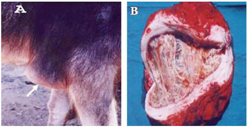
Fig. 1 (A) Olecranon bursitis in a female buffalo. (B) Proliferative form of presternal bursitis in a female buffalo. Note the thin wall and the tissue strands filling the bursal cavity.
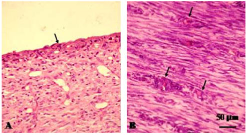
Fig. 2 Histophatological microphotographs of cystic form of presternal bursitis in a male buffalo. (A) Connective tissue-like cells lining the bursal cavity (arrows). (B) Dense connective tissue formed of mature fibrocytes and collagenous fibers. There were also numerous blood capillaries surrounded with inflammatory cells (arrows). H&E strain.

Fig. 3 Histopathological microphotographs of proliferative form of presternal bursitis in a female buffalo. (A) Loss of the cellular lining to more fibrous tissue (asterisk). The underlying connective tissue consisted of mature fibrocytes (arrows) and collagenous fibers. (B) Cross section in connective tissue colloid projected from the wall of bursa into the bursal lumen. Note the connective tissue lining (arrow) and the colloid core (arrowhead). H&E strain.
Reference
-
1. Bancroft JD, Stevans A. Theory and Practice of Histological Techniques. 1990. 3rd ed. Edinburgh: Churchill Livingstone;113–305.2. Davis JM, Broughton SJ. Prepatellar bursitis caused by Brucella abortus. Med J Aust. 1996. 165:460.3. Dietz O. Diseases of the Horse: a Handbook for Science and Practice. Vol. 2. 1984. Berlin: Karger;14–15.4. Honnas CM, Schumacher J, McClure SR, Crabill MR, Carter GK, Schmitz DG, Hoffman AG. Treatment of olecranon bursitis in horses: 10 cases (1986-1993). J Am Vet Med Assoc. 1995. 206:1022–1026.5. Piguet M, Steiner A, Eicher R, Martig J. Surgical treatment of carpal hygroma in cattle: 17 cases (1990-1994). Schweiz Arch Tierheilkd. 1997. 139:210–216.6. Ottaway CA, Worden AN. Bursae and tendon sheaths of the horse. Vet Rec. 1940. 52:477.7. Stashak TS. Stashak TS, editor. Lameness. Adams' Lameness in Horses. 1987. 4th ed. Philadelphia: Lea & Febiger;675.8. Venugopalan A. Essentials of Veterinary Surgery. 1982. 4th ed. New Delhi: Oxford & IBH Publishing;147–165.9. Weiss L. Histology: Cell and Tissue Biology. 1983. 5th ed. New York: Elsevier Biomedical;525.10. Wyn-Jones G. Equine Lameness. 1988. Oxford: Blackwell;120–121.
- Full Text Links
-
- Actions
-
Cited
- CITED
-
- Close
- Share
-
- Similar articles
-
- Osteomyelitis Resulting from Chronic Septic Olecranon Bursitis: Report of Two Cases
- Ultrasound-Guided 50% Ethyl Alcohol Injection for Patients With Malleolar and Olecranon Bursitis: A Prospective Pilot Study
- Surgical Traetment of Ischiogluteal Bursitis: 3 cases report
- Separation of Unfused Olecranon Epiphysis in an Adult Baseball Pitcher: A Case Report
- Fractures of the Olecranon of Ulna treated by Plating and Tension-Band Wiring technique
