Heterosporis anguillarum infections in farm cultured eels (Anguilla japonica) in Korea
- Affiliations
-
- 1National Veterinary Research and Quarantine Service, Ministry of Agriculture and Forestry, Anyang 430-824, Korea. johsj@nvrqs.go.kr
- KMID: 1089665
- DOI: http://doi.org/10.4142/jvs.2007.8.2.147
Abstract
- Ten eels (Anguilla japonica) from a fish farm in Korea were examined and diagnosed with a Heterosporis infection. The gross lesions on the trunk were uneven and the concave parts were pasty. Histopathologically, lyses of the trunk muscles, degenerative muscle fibers and the scattered spores were observed. The sporophorocyst (SPC) contained several spores with a variety of shapes. Some SPC were disrupted and the spores in the SPC were scattered in the muscle tissues. Macrophages existed near the scattered spores. Electron microscopy revealed special structures such as sporophorocyst containing various developmental parasitic stages such as meronts, sporonts, sporophorous vesicles and spores.
MeSH Terms
-
*Anguilla
Animals
Aquaculture
Fish Diseases/*parasitology/pathology
Histocytochemistry/veterinary
Korea
Microscopy, Electron, Scanning Transmission/veterinary
Microsporidia/*growth & development/ultrastructure
Microsporidiosis/parasitology/pathology/*veterinary
Muscular Diseases/parasitology/pathology/*veterinary
Figure
-
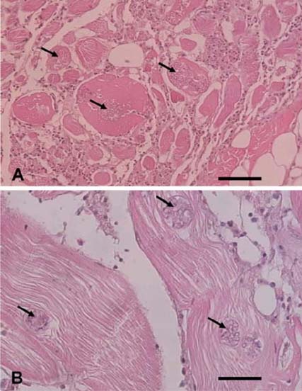
Fig. 1 Optical micrographs of the muscle tissue infected with Heterosporis anguillarum. The tissue was cross-sectioned (A) and sectioned longitudinally (B). Multifocal sporocysts and hyaline degeneration are shown in the muscle fibers (arrowheads) and diffused severe lymphocytic inflammation can be seen in the muscle tissue. bar = 60 µm (A), 30 µm (B).
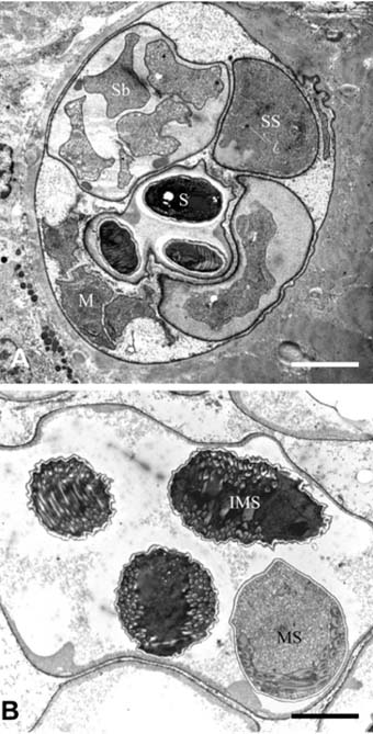
Fig. 2 Transmission electron micrographs of Heterosporis anguillarum. (A) A part of the sporophorocyst (SPC) of Heterosporis anguillarum embedded in the muscle tissue. SPC containing various stages of Heterosporis anguillarum. Presented the meronts (M), sporonts (SS), sporoblasts (Sb) and spore (S) in the sporophorous vesicles. (B) Mature spore (MS) and immature spore (IMS) are shown in the sporophorous vesicle. bar = 2 µm.
Reference
-
1. Canning EU, Nicholas JP. Genus Pleistophora (Phylum Microspora): redescription of the type species, Pleistophora typicalis Gurley, 1893 and ultrastructural characterization of the genus. J Fish Dis. 1980. 3:317–338.
Article2. Hashimoto K, Takinami K. Electron microscopic observations of the spores of Pleistophora anguillarum, a microsporidian parasite of the eel. Bull Jpn Soc Sci Fish. 1976. 42:411–419.
Article3. Hoshina T. On a new microsporidia, Pleistophora anguillarum n.sp. from the muscle of the eel, Anguilla japonica. J Tokyo Univ Fish. 1951. 38:35–49.4. Kano T, Fukui H. Studies on Pleistophora infection in eel, Anguilla japonica. I. Experimental induction of microsporidiosis and fumagillin efficacy. Fish Pathol. 1982. 16:193–200.5. Kano T, Okauchi T, Fukui H. Studies on Pleistophora infection in eel Anguilla japonica. II. Preliminary test for application of fumagillin. Fish Pathol. 1982. 17:107–114.6. Lom J, Dykova I, Wang CH, Lo CF, Kou GH. Ultrastructural justification for the transfer of Pleistophora anguillarum Hoshina, 1959 to the genus Heterosporis Schubert, 1969. Dis Aquat Organ. 2000. 43:225–231.
Article7. Lom J, Dykova I, Tonguthai K. Kabatana gen.n., new name for the microsporidian genus Kabataia Lom, Dykova and Tonguthai. Folia Parasitol. 1999. 47:78.
Article8. Lom J, Nilsen F. Fish microsporidia: fine structural diversity and phylogeny. Int J Parasitol. 2003. 33:107–127.
Article9. Matthews RA, Matthews BF. Cell and tissue reactions of turbot Scophthalmus maximus (L.) to Tetramicra brevifilum gen.n., sp.n (Microspora). J Fish Dis. 1980. 3:495–515.
Article10. Roberts RJ. Parasites of the viscera and musculature. Fish Pathology. 2001. 3rd ed. New York: Harcourt Publishers;285.11. Schubert G. Ultracytologische Untersuchungen an der spore der Mikrosporidienart, Heterosporis finki gen. n., sp. n. Z Parasitenkd. 1969. 32:59–79.12. Suh JW, Chun SK. The infection experiment of Pleistophora to eels, Anguilla japonica and the histopathological investigation of the infection development. Bull Korean Soc Fish Pathol. 1988. 1:51–57.13. Weiss LM. Microsporidia: emerging pathogenic protests. Acta Trop. 2001. 78:89–102.
- Full Text Links
-
- Actions
-
Cited
- CITED
-
- Close
- Share
-
- Similar articles
-
- Korean Mistletoe (Viscum album Coloratum) Extract Induces Eel (Anguilla japonica) Non-specific Immunity
- Positivity and Intensity of Gnathostoma spinigerum Infective Larvae in Farmed and Wild-Caught Swamp Eels in Thailand
- The wormicidal substances of fresh water fishes on Clonorchis sinensis II. Preliminary research on the wormicidal substances from mucous substances of various fresh water fishes
- Occurrence of Gray Mold Caused by Botrytis cinerea on Cryptotaenia japonica in Korea
- Experimental studies on the second intermediate hosts of Clonorchis sinensis. III. Observations on the relationship between clavate cells of epidermis and infectivity of metacercariae of Clonorchis sinensis in fresh-water fish
