Multiple intestinal lymphomatous polyposis in a Jindo dog
- Affiliations
-
- 1Department of Pathology, College of Veterinary Medicine, Kyungpook National University, Daegu 702-701, Korea. jeongks@knu.ac.kr
- 2Dong-In Animal Hospital, Daegu 700-424, Korea.
- KMID: 1089491
- DOI: http://doi.org/10.4142/jvs.2006.7.4.401
Abstract
- A male, 5-year-old Jindo dog underwent enterectomy and enteroanastomosis due to ileus of the intestine at a local veterinary hospital. Grossly, the excised intestine showed markedly thickened multinodular masses in the serosal layer of the upper part, and soft-to-firm, creamcolored neoplastic masses that displayed extensive nodular mucosal protuberances into the lumen. The neoplastic masses were filled with large round cells that were ovoid in shape and they had pale and/or hyperchromatic nuclei. The neoplastic cells had mainly infiltrated into the mucosal and submucosal layers, and they had diffusely invaded the muscular and serosal layers. Therefore, the diagnosis of canine multiple intestinal malignant lymphomatous polyposis was made based on the gross and histopathological findings. The origin of these tumor cells was determined to be B-cells since they were positive for anti-CD20.
MeSH Terms
Figure
-
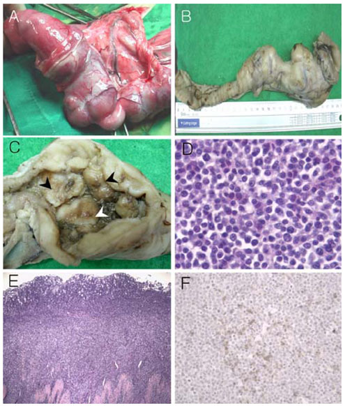
Fig. 1 Gross and histopathological findings of the intestine. (A) Expanded and hypertrophical lesions could be seen in the intestine before enterctomy. (B) The surgically-excised intestine (30 cm in length) included markedly thick multinodular masses in the serosal layer of the upper part and an irregularly thickened wall. (C) Soft-to-firm, cream-colored neoplastic masses (arrowhread) typically showed extensive multi-nodular mucosal protuberances in the lumen. (D) These neoplastic cells were round and ovoid in shape and they were often pale compared to their hyperchromatic nuclei of various sizes with small nucleoli. H&E stain, ×330. (E) These neoplastic cells were mainly infiltrated into mucosal and submucosa, and they had diffusely invaded the muscular and serosal layers. H&E stain, ×33. (F) Tumor cells were positively stained for the CD20 antibody in the tumorous lesions. Immunostaining for CD20 antibody, ×132.
Reference
-
1. Coyle KA, Steinberg H. Characterization of lymphocytes in canine gastrointestinal lymphoma. Vet Pathol. 2004. 41:141–146.
Article2. Hirakawa K, Fuchigami T, Nakamura S, Daimaru Y, Ohshima K, Sakai Y, Ichimaru T. Primary gastrointestinal T-cell lymphoma resembling multiple lymphomatous polyposis. Gastroenterology. 1996. 111:778–782.
Article3. Isaacson PG, MacLennan KA, Subbuswamy SG. Multiple lymphomatous polyposis of the gastrointestinal tract. Histopathology. 1984. 8:641–656.
Article4. Jubala CM, Wojcieszyn JW, Valli VE, Getzy DM, Fosmire SP, Coffey D, Bellgrau D, Modiano JF. CD20 Expression in Normal Canine B Cells and in Canine non-Hodgkin Lymphoma. Vet Pathol. 2005. 42:468–476.
Article5. Jubb KVF, Kennedy PC, Palmer N. Pathology of Domestic Animals. Vol. 2. 1993. San Diego: Academic Press;140.6. Kadayifci A, Benekli M, Savas MC, Arslan S, Uzunalimoglu B, Barista I, Gullu IH, Tekuzman G. Multiple lymphomatous polyposis. J Surg Oncol. 1997. 64:336–340.
Article7. Lavergne A, Brouland JP, Launay E, Nemeth J, Ruskone-Fourmestraux A, Galian A. Multiple lymphomatous polyposis of the gastrointestinal tract. An extensive histopathologic and immunohistochemical study of 12 cases. Cancer. 1994. 74:3042–3050.
Article8. Moore PF, Vernau W. Feldman BF, Zinkl JG, Jain NC, editors. Lymphocytes: differentiation molecules in diagnosis and prognosis. Schalm's Veterinary Hematology. 2000. 5th ed. Philadelphia: Lippincott Williams & Wilkins;247–255.9. Sheahan DG, Martin F, Baginsky S, Mallory GK, Zamcheck N. Multiple lymphomatous polyposis of the gastrointestinal tract. Cancer. 1971. 28:408–425.
Article10. von Schilling C. Immunotherapy with anti-CD20 compounds. Semin Cancer Biol. 2003. 13:211–222.
Article
- Full Text Links
-
- Actions
-
Cited
- CITED
-
- Close
- Share
-
- Similar articles
-
- Multiple lymphomatous polyposis of the gastrointestinal tract: a report of two cases with immunohistochemical studies
- Multiple Lymphomatous Polyposis of the Gastrointestinal Tract: Report of Three Cases
- A Case of Recurrent Mantle Cell Lymphoma in the form of Intestinal Lymphomatous Polyposis
- A Case of Multiple Lymphomatous Polyposis
- A Case of Synchronous Gastrointestinal Multiple Lymphomatous Polyposis and Gastric Adenocarcinoma
