Korean J Radiol.
2009 Apr;10(2):121-128. 10.3348/kjr.2009.10.2.121.
Necrotizing Fasciitis versus Pyomyositis: Discrimination with Using MR Imaging
- Affiliations
-
- 1Department of Radiology, The Catholic University of Korea, Korea. whjee@catholic.ac.kr
- 2Department of Pathology, The Catholic University of Korea, Korea.
- 3Department of Internal Medicine, The Catholic University of Korea, Korea.
- 4Department of Orthopedic Surgery, The Catholic University of Korea, Korea.
- 5Department of Internal Medicine, Kyung Hee University, Korea.
- KMID: 1088722
- DOI: http://doi.org/10.3348/kjr.2009.10.2.121
Abstract
OBJECTIVE
We wanted to evaluate the MR findings for differentiating between necrotizing fasciitis (NF) and pyomyositis (PM).
MATERIALS AND METHODS
The MR images of 19 patients with surgically confirmed NF (n = 11) and pathologically confirmed PM (n = 8) were retrospectively reviewed with regard to the presence or absence of any MRI finding criteria that could differentiate between them.
RESULTS
The patients with NF had a significantly greater prevalence of the following MR findings (p < 0.05): a peripheral band-like hyperintense signal in muscles on fat-suppressed T2-weighted images (73% of the patients with NF vs. 0% of the patients with PM), peripheral band-like contrast enhancement (CE) of muscles (82% vs. 0%, respectively) and thin smooth enhancement of the deep fascia (82% vs. 13%, respectively). The patients with PM had a significantly greater prevalence of the following MRI findings (p < 0.05): a diffuse hyperintense signal in muscles on fat-suppressed T2-weighted images (27% of the patients with NF vs. 100% in the patients with PM), diffuse CE of muscles (18% vs. 100%, respectively), thick irregular enhancement of the deep fascia (0% vs. 75%, respectively) and intramuscular abscess (0% vs. 88%, respectively). For all patients with NF and PM, the superficial fascia and muscle showed hyperintense signals on T2-weighted images and CE was seen on fat-suppressed CE T1-weighted images. The subcutaneous tissue and deep fascia showed hyperintense signals on T2-weighted images and CE was seen in all the patients with NF and in seven (88%) of the eight patients with PM, respectively.
CONCLUSION
MR imaging is helpful for differentiating between NF and PM.
Keyword
MeSH Terms
Figure
-
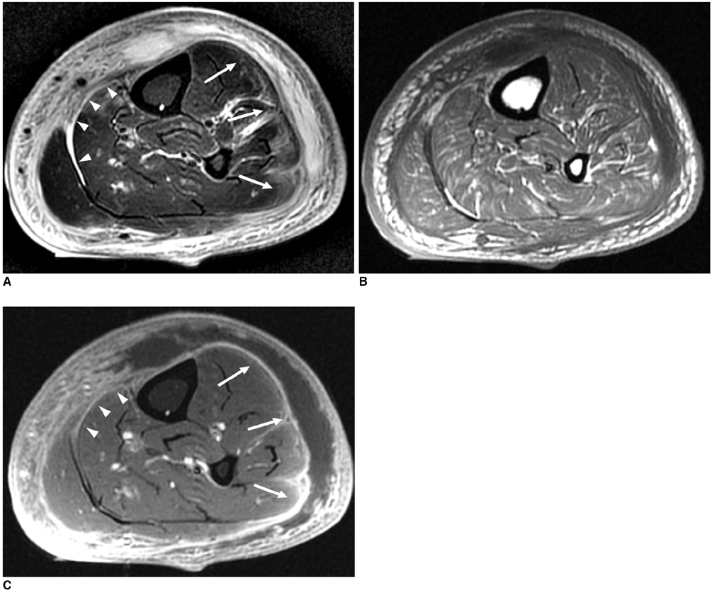
Fig. 1 51-year-old man with surgically confirmed necrotizing fasciitis.
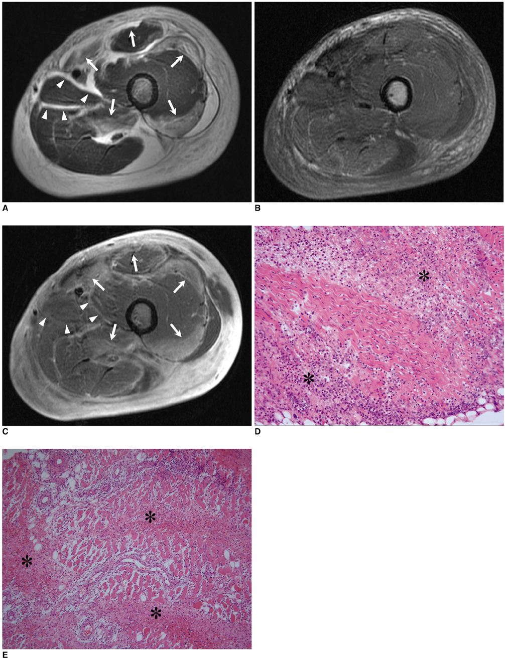
Fig. 2 37-year-old man with surgically confirmed necrotizing fasciitis.
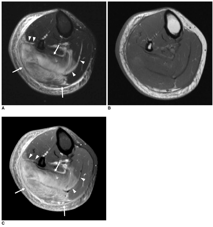
Fig. 3 29-year-old woman with pathologically confirmed pyomyositis.
Cited by 2 articles
-
Clinical Practice Guidelines for Soft Tissue Infections
, , , ,
Infect Chemother. 2012;44(4):213-232. doi: 10.3947/ic.2012.44.4.213.Two Cases of Necrotizing Fasciitis in Patients with SLE
Dong-su Shin, Mi-ryeong Seo, Hyung-jeong Cho, Hyo-jin Choi, Eun-bong Lee, Han-joo Baek
J Rheum Dis. 2011;18(2):132-136. doi: 10.4078/jrd.2011.18.2.132.
Reference
-
1. Wilson B. Necrotizing fasciitis. Am J Surg. 1952. 18:416–431.2. Brothers TE, Tagge DU, Stutley JE, Conway WF, Del Schutte H Jr, Byrne TK. Magnetic resonance imaging differentiates between necrotizing and non-necrotizing fasciitis of the lower extremity. J Am Coll Surg. 1998. 187:416–421.3. Arslan A, Pierre-Jerome C, Borthne A. Necrotizing fasciitis: unreliable MRI findings in the preoperative diagnosis. Eur J Radiol. 2000. 36:139–143.4. McAllister L. Kransdorf MJ, Murphey MD, editors. Masses that may mimic soft tissue tumors. Imaging of soft tissue tumors. 2006. 2nd ed. Philadelphia: Lippincott Williams & Wilkins;511–572.5. Wall DB, de Virgilio C, Black S, Klein SR. Objective criteria may assist in distinguishing necrotizing fasciitis from nonnecrotizing soft tissue infection. Am J Surg. 2000. 179:17–21.6. Beauchamp NJ Jr, Scott WW Jr, Gottlieb LM, Fishman EK. CT evaluation of soft tissue and muscle infection and inflammation: a systematic compartmental approach. Skeletal Radiol. 1995. 24:317–324.7. Schmid MR, Kossmann T, Duewell S. Differentiation of necrotizing fasciitis and cellulitis using MR imaging. AJR Am J Roentgenol. 1998. 170:615–620.8. Duewell S, Hagspiel KD, Zuber J, von Schulthess GK, Bollinger A, Fuchs WA. Swollen lower extremity: role of MR imaging. Radiology. 1992. 184:227–231.9. Revelon G, Rahmouni A, Jazaerli N, Godeau B, Chosidow O, Authier J, et al. Acute swelling of the limbs: magnetic resonance pictorial review of fascial and muscle signal changes. Eur J Radiol. 1999. 30:11–21.10. Martin DR, Single LA. Molecular epidemiology of group A streptococcus M type 1 infections. J Infect Dis. 1993. 167:1112–1117.11. Bisno AL, Stevens DL. Streptococcal infections of skin and soft tissues. N Engl J Med. 1996. 334:240–245.12. Gordon BA, Martinez S, Collins AJ. Pyomyositis: characteristics at CT and MR imaging. Radiology. 1995. 197:279–286.13. Stamenkovic I, Lew PD. Early recognition of potentially fatal necrotizing fasciitis. The use of frozen-section biopsy. N Engl J Med. 1984. 310:1689–1693.14. Rahmouni A, Chosidow O, Marthieu D, Gueorguieva E, Jazaerli N, Radier C, et al. MR imaging in acute infectious cellulitis. Radiology. 1994. 192:493–496.15. Struk DW, Munk PL, Lee MJ, Ho SG, Worsley DF. Imaging of soft tissue infections. Radiol Clin North Am. 2001. 39:277–303.16. Fugitt JB, Puckett ML, Quigley MM, Kerr SM. Necrotizing fasciitis. Radiographics. 2004. 24:1472–1476.17. Loh NN, Ch'en IY, Cheung LP, Li KC. Deep fascial hyperintensity in soft-tissue abnormalities as revealed by T2-weighted MR imaging. AJR Am J Roentgenol. 1997. 168:1301–1304.
