Yonsei Med J.
2010 May;51(3):463-465. 10.3349/ymj.2010.51.3.463.
Laparoscopic Treatment of Appendicovesical Fistula
- Affiliations
-
- 1Department of General Surgery, Bundang CHA Hospital, CHA University College of Medicine, Seongnam, Korea.
- 2Department of Radiology, Bundang CHA Hospital, CHA University College of Medicine, Seongnam, Korea.
- 3Department of Urology, Yonsei Medical Center, Yonsei University College of Medicine, Seoul, Korea.
- 4Department of Urology, Bundang CHA Hospital, CHA University College of Medicine, Seongnam, Korea. urohong@yahoo.co.kr
- KMID: 1075004
- DOI: http://doi.org/10.3349/ymj.2010.51.3.463
Abstract
- A 23-year-old man had a history of intermittent episodes of urinary tract infection with associated low abdominal pain for 15 years. Persistent bacteriuria even with prolonged antibiotics was the reason why he was referred to our hospital. Laboratory tests were normal except pyuria and growth of Escherichia coli in the urinary samples. Cystoscopy revealed a small slit-like opening on the right lateral wall of bladder dome. We found some air within the bladder and a suspicious communicating tract between the appendix and bladder on a CT scan. With a strong impression of appendicovesical fistula, a laparoscopy was performed to confirm a diagnosis and to remove the appendicovesical fistula resulting in a satisfactory result without any complication.
MeSH Terms
Figure
-

Fig. 1 Cystoscopic finding shows a slit-like opening (arrowhead) on the right side of the bladder dome with a fecalith (arrow).

Fig. 2 Protrusion of right side of the bladder dome on voiding cystourethrography.
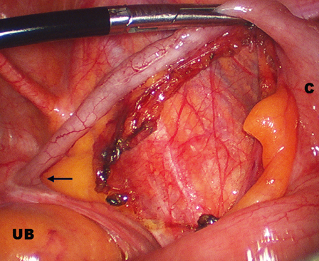
Fig. 3 Laparoscopic finding shows a grasped appendix, of which the tip is firmly attached to the right side of the bladder dome (arrow). UB, urinary bladder; C, cecum.
Cited by 1 articles
-
Vesico-Appendiceal Fistula Misdiagnosed as Meckel's Diverticulum: A Laparoscopic Approach
Jung Woo Lee, Jae Hyun Ahn, Hong Koo Ha
World J Mens Health. 2012;30(3):195-197. doi: 10.5534/wjmh.2012.30.3.195.
Reference
-
1. Steel MC, Jones IT, Webb D. Appendicovesical fistula arising from appendiceal diverticulum suspected on barium enema. ANZ J Surg. 2001. 71:769–770.
Article2. Abubakar AM, Pindiga UH, Chinda JY, Nggada HA. Appendicovesical fistula associated with Hirschsprung's disease. Pediatr Surg Int. 2006. 22:617–618.3. Athanassopoulos A, Speakman MJ. Appendicovesical fistula. Int Urol Nephrol. 1995. 27:705–708.4. Bigler ME, Wofford JE, Pratt SM, Stone WJ. Serendipitous diagnosis of appendicovesical fistula by bone scan: a case report. J Urol. 1989. 142:815–816.
Article5. Goldman SM, Fishman EK, Gatewood OM, Jones B, Siegelman SS. CT in the diagnosis of enterovesical fistulae. AJR Am J Roentgenol. 1985. 144:1229–1233.6. Sarr MG, Fishman EK, Goldman SM, Siegelman SS, Cameron JL. Enterovesical fistula. Surg Gynecol Obstet. 1987. 164:41–48.7. Gross M, Peng B. Appendico-vesical fistula. J Urol. 1969. 102:697–698.8. Albrecht K, Schumann R, Peitgen K, Walz MK. [Laparoscopic therapy of appendicovesical fistula -- two case reports.]. Zentralbl Chir. 2004. 129:396–398.9. Afifi AY, Fusia TJ, Feucht K, Paluzzi MW. Laparoscopic treatment of appendicovesical fistula: a case report. Surg Laparosc Endosc. 1994. 4:320–324.
- Full Text Links
-
- Actions
-
Cited
- CITED
-
- Close
- Share
-
- Similar articles
-
- A fecalith mimicking a bladder calculus secondary to an appendicovesical fistula: a case report
- Laparoscopic Repair for Enterocutaneous Fistula Caused by Laparoscopic Right Hemicolectomy for Pan-Peritonitis due to Cecal Cancer Perforation
- Case Report: Gastrobronchial Fistula after Sleeve Gastrectomy: Treated by Laparoscopic Proximal Gastrectomy with Double Tract Reconstruction
- Incidental cholecystojejunal fistula treated with successful laparoscopic management
- Laparoscopic Management of Vesicouterine Fistula due to Intrauterine Device
