Multidetector CT Urography in Imaging of the Urinary Tract in Patients with Hematuria
- Affiliations
-
- 1Department of Radiology, Massachusetts General Hospital, Harvard Medical School. ssaini@partners.org
- KMID: 1066239
- DOI: http://doi.org/10.3348/kjr.2004.5.1.1
Abstract
- This review article comprehensively discusses multidetector CT urography protocols and their role in imaging of the urinary tract in patients with hematuria.
Keyword
MeSH Terms
Figure
-
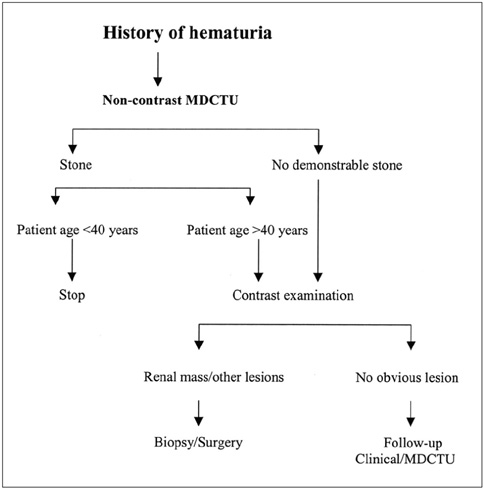
Fig. 1 A proposed algorithm for imaging of patients with hematuria.

Fig. 2 Multidetector CT urography in a 36 year-old woman with hematuria. Nephropyelographic phase axial multidetector CT urography image (A) shows an obstructed upper pole moiety which is elegantly confirmed on a coronal multidetector CT urography image (B).

Fig. 3 Multidetector CT urography examination of a 65-year-old lady with left renal caliceal diverticulum and nodular calcification. Supine (A) and prone images (B) show change in position of this nodular calcific density (arrows) suggestive of calculus.
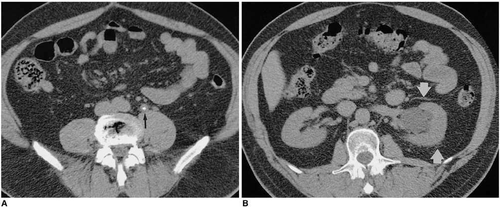
Fig. 4 Unenhanced multidetector CT urography in a 46-year-old man with hematuria and colicky pain in left flank demonstrates left mid-ureteral calculus (A) with "tissue-rim sign" (arrow) and proximal hydronephrosis (B) with minimal perinephric fat stranding (arrows).

Fig. 5 Multidetector CT urography study in a 52-year-old lady hematuria demonstrating renal cell carcinoma. Unenhanced image (A) demonstrates right renal mass with calcification. Contrast-enhanced images demonstrate a hypervascular enhancing mass (B).
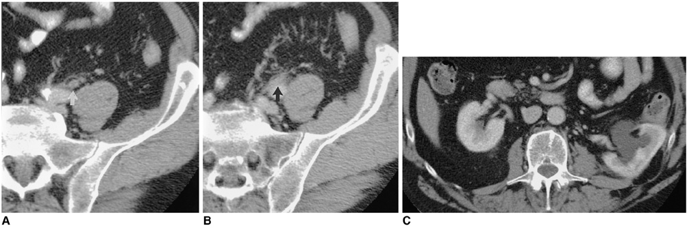
Fig. 6 Multidetector CT urography image of a 53-year-old man with hematuria secondary to a transitional cell carcinoma of left ureter shows thickened, enhancing ureteric wall (arrow) with periureteric fat stranding suggestive of urothelial lesion with extramural spread of the disease (A and B). Proximal hydronephrosis with hydroureter is noted (C).

Fig. 7 A 42-year-old man underwent multidetector CT urography examination for hematuria. An eccentric filling defect (arrow) in the distal left ureter was reported as suspicious for urothelial neoplasm on axial post-contrast (A), coronal (B) and sagittal (C) maximum intensity projection images. Retrograde pyelography did not reveal any intraluminal abnormality. In retrospect, the "filling defect" was thought to be due to vascular impression.

Fig. 8 Multidetector CT urography evaluation of a 71-year-old lady with macroscopic hematuria secondary to transitional cell carcinoma of urinary bladder. A large polypoidal mass is seen arising from the right lateral wall with subtle perivesical fat stranding (arrows) suggestive of extramural spread.

Fig. 9 Multidetector CT urography study in a 75-year-old man with uncontrolled diabetes mellitus, left flank pain, hematuria and fever. A large staghorn calculus (arrow) with hypodense, non-enhancing parenchymal mass is seen in the left kidney suggestive of xanthogranulomatous pyelonephritis.
Reference
-
1. Chow LC, Sommer FG. Multidetector CT urography with abdominal compression and three-dimensional reconstruction. AJR Am J Roentgenol. 2001. 177:849–855.2. Kim JK, Ahn JH, Park T, Ahn HJ, Kim KS, Cho KS. Virtual cystoscopy of the contrast-filled bladder in patients with gross hematuria. AJR Am J Roentgenol. 2002. 179:763–768.3. Pollack HM. Gagliardi RA, McClennan BL, editors. Uroradiology. A history of the radiological science. 1996. Reston, Virginia: American Roentgen Ray Society;237–238.4. Amis ES. Epitaph for the urogram. Radiology. 1999. 213:639–640.5. Fielding JR, Silver SG, Rubin GD. Helical CT of the urinary tract. AJR Am J Roentgenol. 1999. 172:1199–1206.6. Urban BA. The small mass: What is the role of multiphasic helical scanning? Radiology. 1997. 202:22–23.7. Chai RY, Jhaveri K, Saini S, Hahn PF, Nichols S, Mueller PR. Comprehensive evaluation of patients with hematuria on a multislice computed tomography scanner: Protocol designs and preliminary observations. Australas Radiol. 2001. 45:536–539.8. Grossfield GD, Litwin MS, Wolf JS, et al. Evaluation of asymptomatic microscopic hematuria in adults. The American Urological Association Best Practice Policy-Part 1: definition, detection prevalence and etiology. Urology. 2001. 57:599–603.9. Jones DL, Langstaff RJ, Holy SD, et al. The value of cystoureteroscopy in the investigation of microscopic hematuria: a prospective study of 100 patients. Br J Urol. 1988. 62:541–545.10. Grossfield GD, Litwin MS, Wolf JS, et al. Evaluation of asymptomatic microscopic hematuria in adults. The American Urological Association Best Practice Policy-Part II: Patient evaluation, cytology, voided markers, imaging, cystsocopy, nephrology, evaluation and follow-up. Urology. 2001. 57:604–610.11. Maher MM, Jhaveri K, Lucey BC, et al. Does the administration of saline flush during CT urography (CTU) improve ureteric distension and opacification? Radiology. 2000. 221(p):500.12. McTavish JD, Jinaki M, Zou KH, Silverman SG. Multidetector CT urography: analysis of techniques and comparison with IVU. Radiology. 2000. 27(p):225.13. Caoili EM, Cohan RH, Korobkin M, et al. Urinary Tract Abnormalities: Initial Experience with Multi-Detector Row CT Urography. Radiology. 2002. 222:353–360.14. Heneghan JP, Kim DH, Leder RA, DeLong D, Nelson RC. Compression CT urography: a comparison with IVU in the opacification of the collecting system and ureters. J Comput Assist Tomogr. 2001. 25:343–347.15. Birnbaum BA, Jacobs JE, Ramchandani P. Multiphasic renal CT: comparison of renal mass enhancement during the corticomedullary and nephrographic phases. Radiology. 1996. 200:753–758.16. Sussman SK, Illescas FF, Opalacz JP, et al. Renal streak artifact during contrast enhanced CT. comparison of high versus low osmolality contrast media. Abdom Imaging. 1993. 18:180–185.17. Maher MM, Prassad TA, Fitzpatrick JM, et al. Spinal dysraphism at MR Urography: Initial experience. Radiology. 2000. 216:237–241.18. Kim JK, Cho KS. CT urography and virtual endoscopy: promising imaging modalities for urinary tract evaluation. Br J Radiol. 2003. 76:199–209.19. Levine JA, Neitlich J, Verga M, et al. Ureteral calculi in patients with flank pain: correlation of plain radiography with unenhanced helical CT. Radiology. 1997. 204:27–31.20. Fielding JR, Fox LA, Heller H, et al. Spiral CT in the evaluation of flank pain: overall accuracy and feature analysis. J Comput Assist Tomogr. 1997. 21:635–658.21. Dalla Palma L. What is left of IV urography? Eur Radiol. 2001. 11:931–939.22. Kawashima A, Sandler CM, Boridy IC, et al. Unenhanced helical CT of ureterolithiasis: value of the tissue rim sign. AJR Am J Roentgenol. 1997. 168:997–1000.23. Bell TV, Fenlon HM, Davison BD, et al. Unenhanced helical CT criteria to differentiate distal ureteral calculi from pelvic phleboliths. Radiology. 1998. 207:363–367.24. Boridy IC, Nikolaidis P, Kawashima A, et al. Ureterolithiasis: value of the tail sign in differentiating phleboliths from ureteral calculi at nonenhanced helical CT. Radiology. 1999. 211:619–621.25. Coll DM, Varanelli MJ, Smith RC. Relationship of spontaneous passage of ureteral calculi to stone size and location as revealed by unenhanced helical CT. AJR Am J Roentgenol. 2002. 178:101–103.26. Jamis-Dow CA, Choyke PL, Jennings SB, et al. Small (<3 cm) renal masses: detection with CT versus US and pathologic correlation. Radiology. 1996. 198:785–788.27. Polascik TJ, Pound CR, Meng MV, et al. Partial nephrectomy: technique, complications and pathological findings. J Urol. 1995. 154:1312–1318.28. Polascik TJ, Meng MV, Epstein JI, et al. Intraoperative sonography for the evaluation and management of renal tumors: experience with 100 patients. J Urol. 1995. 154:1676–1680.29. Bosniak MA. The current radiological approach to renal cysts. Radiology. 1986. 158:1–10.30. Abdulla C, Kalra MK, Saini S, et al. Pseudoenhancement of "renal cysts" by different multidetector CT scanners: a phantom experiment. AJR Am J Roentgenol. 2002. 179:1473–1476.31. Bosniak MA, Rofsky NM. Problems in the detection and characterization of small renal masses. Radiology. 1996. 198:638–641.32. Liu W, Esler SJ, Kenny BJ, et al. Low-dose nonenhanced helical CT of renal colic: assessment of ureteric stone detection and measurement of effective dose equivalent. Radiology. 2000. 215:51–55.33. Atasoy C, Yagci C, Fitoz S, et al. Cross-sectional imaging in ureter tumors: findings and staging. Clin Imaging. 2001. 25:197–202.34. Song JH, Francis IR, Platt JF, et al. Bladder tumor extension at virtual cystoscopy. Radiology. 2001. 218:95–100.35. Kwashima A, LeRoy AJ. Radiologic evaluation of patients with renal infections. Infect Dis Clin North Am. 2003. 17:433–456.36. Kawashima A, Sandler CM, Goldman SM, et al. CT of renal inflammatory disease. RadioGraphics. 1997. 17:851–866.37. Kawashima A, Sandler CM, Goldman SM. Current roles and controversies in the imaging evaluation of acute renal infection. World J Urol. 1998. 16:9–17.38. Kawashima A, Fishman EK, Goldman SM, et al. Helical CT of acute pyelonephritis: is there an ideal timing for imaging? Radiology. 1996. 201:148–149.39. Hoddick W, Jeffrey RB, Goldberg HI, et al. CT and sonography of severe renal and perirenal infections. AJR Am J Roentgenol. 1983. 140:517–520.40. Lang EK, Macchia RJ, Thomas R, et al. Detection of medullary and papillary necrosis at an early stage multiphase helical computerized tomography. J Urol. 2003. 170:94–98.
- Full Text Links
-
- Actions
-
Cited
- CITED
-
- Close
- Share
-
- Similar articles
-
- Upper Urinary Tract Gross Hematuria: Clinical Diagnosis and Disease Distribution
- Comparison of CT Urography and Intravenous Urography in Patients with Hematuria
- Drug-induced MR Urography: The effects of Furosemide and Intravenous Saline Injection on MR Urography of Obstructed and Non-obstructed Urinary Tract
- A Clinical Observation on Urothelial Tumor of the Upper Urinary Tract
- Clinical Usefulness of Multi-Detector Computed Tomography in Diagnosis of Patients with Microscopic Hematuria
