Application of ventriculoperitoneal shunt as a treatment for hydrocephalus in a dog with syringomyelia and Chiari I malformation
- Affiliations
-
- 1The United Graduate School of Veterinary Sciences, Yamaguchi University, 1677-1, Yoshida, Yamaguchi 753-8515, Japan.
- 2Departments of Veterinary Surgery, Faculty of Agriculture, Yamaguchi University, 1677-1, Yoshida, Yamaguchi 753-8515, Japan. b3646@yamaguchi-u.ac.jp
- 3Departments of Veterinary Internal Medicine, Faculty of Agriculture, Yamaguchi University, 1677-1, Yoshida, Yamaguchi 753-8515, Japan.
- KMID: 1059210
- DOI: http://doi.org/10.4142/jvs.2006.7.2.203
Abstract
- A twenty-month-old Chihuahua male dog was presented to us suffering with ataxia. Based on the physical examination, X-ray and magnetic resonance imaging (MRI) examinations, we diagnosed the dog with hydrocephalus, Chiari I malformation and syringomyelia. Treatment consisted of internal medical treatment and the placement of a ventriculoperitoneal (VP) shunt. The ventricular dilatation was relieved and the dog improved neurologically; however, the Chiari I malformation and syringomyelia remained after surgically positioning the VP shunt.
MeSH Terms
Figure
-

Fig. 1 Lateral radiography reveals a mildly enlarged skull, an open fontanelle and a partial protrusion on the occipital bone.
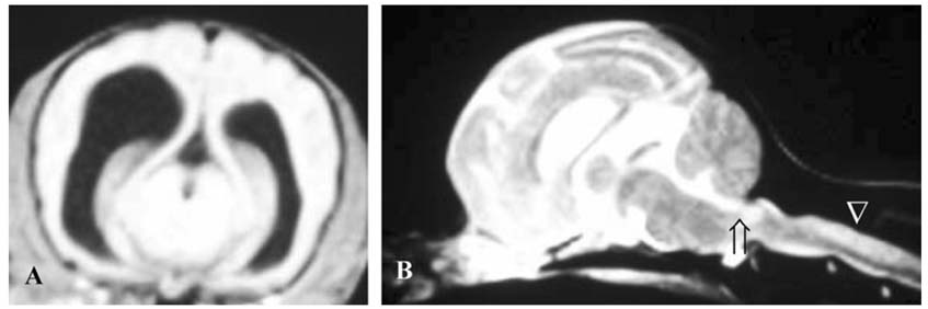
Fig. 2 T1-weighed transverse (A) and T2-weighed sagittal (B) MRI scans, demonstrating asymmetrically enlarged lateral ventricles, slight dilation of the third ventricle, syringomyelia in the region of C2, 3, 4 (B, arrowhead) and caudal (foramen magnum) descent of the cerebellum (Chiari I malformation) (B, arrow).

Fig. 3 The VP shunt was placed into the left lateral ventricle (arrow) and the peritoneal cavity (arrowhead).
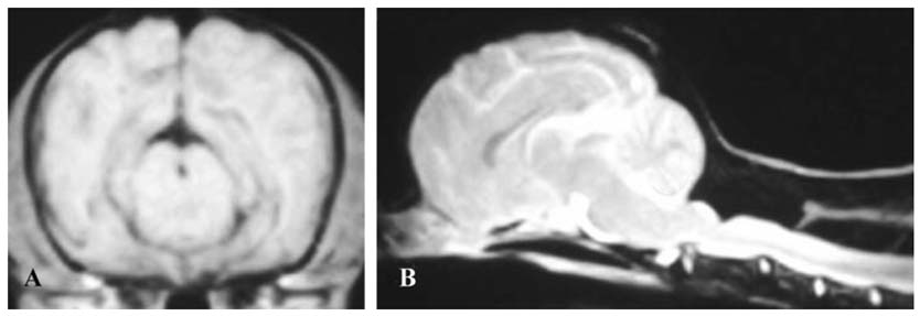
Fig. 4 T1-weighed transverse (A) and T2-weighed sagittal (B) MRI scans 4 months from the date of surgery, demonstrating the relief of dilation of ventricles but the continued presence of Chiari I malformation and syringomyelia (B).
Reference
-
1. Bagley RS. Slatter D, editor. Intracranial surgery. Textbook of Small Animal Surgery. 2003. 3rd ed. Philadelphia: Saunders;1261–1277.2. Eule JM, Erickson MA, O'Brien MF, Handler M. Chiari I malformation associated with syringomyelia and scoliosis: a twenty-year review of surgical nonsurgical treatment in a pediatric population. Spine. 2002. 27:1451–1455.
Article3. Fenner WR. Ettinger SJ, Feldman EC, editors. Diseases of the brain. Textbook of Veterinary Internal Medicine. 1995. 4th ed. Philadelphia: Saunders;578–629.4. Harrington ML, Bagley RS, Moore MP. Hydrocephalus. Vet Clin North Am Small Anim Pract. 1996. 26:843–856.
Article5. Hoppe-Hirsch E, Sainte RC, Renier D, Hirsch JF. Pericerebral collections after shunting. Childs Nerv Syst. 1987. 3:97–102.
Article6. Kang J, Lee IW. Long-term follow-up of shunting therapy. Childs Nerv Syst. 1999. 15:711–717.
Article7. Kitagawa M, Kanayama K, Sakai T. Subdural accumulation of fluid in a dog after the insertion of a ventriculoperitoneal shunt. Vet Rec. 2005. 156:206–208.
Article8. Klekamap J. The pathophysiology of syringomyelia-historical overview and current concept. Acta Neurochir (Wien). 2002. 144:649–664.9. Milhorat TH, Chou MW, Trinidad EM, Kula RW, Mandell M, Wolpert C, Speer MC. Chiari I malformation redefined: clinical and radiographic findings for 364 symptomatic patients. Neurosurgery. 1999. 44:1005–1017.
Article10. Platt SR, Olby NJ. BSAVA Manual of Canine and Feline Neurology. 2004. 3rd ed. Gloucester: British Small Animal Veterinary Association;120–123.11. Pudenz RH, Foltz EL. Hydrocephalus: overdrainage by ventricular shunts. A review and recommendations. Surg Neurol. 1991. 35:200–212.
Article
- Full Text Links
-
- Actions
-
Cited
- CITED
-
- Close
- Share
-
- Similar articles
-
- Acquired Chiari-malformation after Ventriculoperitoneal Shunt for Hydrocepalus Associated with Neurocysticercosis
- Co-Occurrence of Syringomyelia and Hydrocephalus in a Patient Without Chiari Malformation in Her 50's
- Presyrinx Associated with Post-Traumatic Hydrocephalus Successfully Treated by Ventriculoperitoneal Shunt
- Treatment of Syringomyelia due to Chiari Type I Malformation with Syringo-Subarachnoid-Peritoneal Shunt
- Chiari 1.5 Malformation : An Advanced Form of Chiari I Malformation
