Korean J Ophthalmol.
2011 Aug;25(4):285-288. 10.3341/kjo.2011.25.4.285.
Late-Onset Citrobacter koseri Endophthalmitis with Suture Exposure after Secondary Intraocular Lens Implantation
- Affiliations
-
- 1Department of Ophthalmology, Yonsei University Medical Center, Seoul, Korea.
- 2Department of Ophthalmology, National Health Insurance Corporation Ilsan Hospital, Goyang, Korea. eunjee95@hanmail.net
- KMID: 1018420
- DOI: http://doi.org/10.3341/kjo.2011.25.4.285
Abstract
- A 54-year-old male patient was seen in clinic for ocular pain and decreased vision in the right eye with duration of two days. He underwent a cataract operation for his right eye 12 years ago, then a sclera-fixated secondary intraocular implantation and pars plana vitrectomy three years ago due to intraocular lens dislocation. At the initial visit, his visual acuity was restricted to the perception of hand motion. An edematous cornea, cells, flare with hypopyon, and exposed suture material at were observed at the six o'clock direction by slit lamp. Vitreous opacity was noted from B-scan ultrasonography. The patient was diagnosed with late-onset endophthalmitis and an intravitreal cocktail injection was done. On the next day, the hypopyon was aggravated, and therefore a pars plana vitrectomy was performed. A vitreous culture tested positive for Citrobacter koseri. After 12 weeks, the best corrected visual acuity of the right eye improved to 0.7 and a fundus examination revealed a relatively normal optic disc and retinal vasculature. We herein report the first case of endophthalmitis caused by Citrobacter koseri in Korea. Exposed suture material was suspected as the source of infection in this case and prompt surgical intervention resulted in a relatively good visual outcome.
MeSH Terms
-
Anti-Bacterial Agents/administration & dosage
Cataract Extraction/adverse effects
Citrobacter koseri/*isolation & purification
Diagnosis, Differential
Endophthalmitis/diagnosis/*microbiology/therapy
Enterobacteriaceae Infections/diagnosis/*microbiology/therapy
Eye Infections, Bacterial/diagnosis/*microbiology/therapy
Follow-Up Studies
Humans
Intravitreal Injections
Lens Implantation, Intraocular/*adverse effects
Male
Microscopy, Acoustic
Middle Aged
Surgical Wound Infection/diagnosis/*microbiology/therapy
Sutures/adverse effects/microbiology
Visual Acuity
Vitrectomy
Vitreous Body/*microbiology
Figure
-
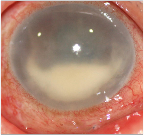
Fig. 1 Slit lamp photography of the right eye at initial presentation. Severe conjunctival injection and exposed suture material at 6 o'clock direction were observed. Also, an edematous cornea with 3.0-mm-height hypopyon was observed.
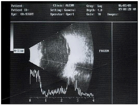
Fig. 2 B-scan ultrasonography of the right eye at initial presentation. Significant vitreous opacity was observed.
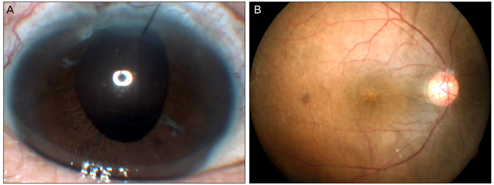
Fig. 3 Twelve weeks after vitrectomy, best corrected visual acuity was improved to 0.7 (refractive errors +12.0 sph -1.50 cyl Ax90), according to a Snellen chart examination. (A) Cornea and anterior chamber were both clear. (B) According to a fundus examination, a relatively normal optic disc and retinal vasculature were observed.
Reference
-
1. Driebe WT Jr, Mandelbaum S, Forster RK, et al. Pseudophakic endophthalmitis. Diagnosis and management. Ophthalmology. 1986. 93:442–448.2. Results of the Endophthalmitis Vitrectomy Study. A randomized trial of immediate vitrectomy and of intravenous antibiotics for the treatment of postoperative bacterial endophthalmitis Endophthalmitis Vitrectomy Study Group. Arch Ophthalmol. 1995. 113:1479–1496.3. Microbiologic factors and visual outcome in the endophthalmitis vitrectomy study. Am J Ophthalmol. 1996. 122:830–846.4. Olson JC, Flynn HW Jr, Forster RK, Culbertson WW. Results in the treatment of postoperative endophthalmitis. Ophthalmology. 1983. 90:692–699.5. Essex RW, Charles PG, Allen PJ. Three cases of post-traumatic endophthalmitis caused by unusual bacteria. Clin Experiment Ophthalmol. 2004. 32:445–447.6. Insler MS, Kook MS, Mani H, Peyman GA. Citrobacter diversus endophthalmitis following penetrating keratoplasty. Am J Ophthalmol. 1988. 106:632–633.7. Hykin PG, Tobal K, McIntyre G, et al. The diagnosis of delayed post-operative endophthalmitis by polymerase chain reaction of bacterial DNA in vitreous samples. J Med Microbiol. 1994. 40:408–415.8. Chen KJ, Sun MH, Hwang YS, et al. Endophthalmitis caused by Citrobacter species. Ocul Immunol Inflamm. 2008. 16:147–153.9. Lalwani GA, Flynn HW Jr, Scott IU, et al. Acute-onset endophthalmitis after clear corneal cataract surgery (1996-2005). Clinical features, causative organisms, and visual acuity outcomes. Ophthalmology. 2008. 115:473–476.10. Doft BH, Kelsey SF, Wisniewski S, et al. Treatment of endophthalmitis after cataract extraction. Retina. 1994. 14:297–304.11. Scott IU, Flynn HW Jr, Feuer W. Endophthalmitis after secondary intraocular lens implantation. A case-report study. Ophthalmology. 1995. 102:1925–1931.12. Kattan HM, Flynn HW Jr, Pflugfelder SC, et al. Nosocomial endophthalmitis survey. Current incidence of infection after intraocular surgery. Ophthalmology. 1991. 98:227–238.13. Leopold IH. Management of intra-ocular infection. Trans Ophthalmol Soc U K. 1971. 91:577–610.14. Altan T, Acar N, Kapran Z, et al. Acute-onset endophthalmitis after cataract surgery: success of initial therapy, visual outcomes, and related factors. Retina. 2009. 29:606–612.15. Somani S, Grinbaum A, Slomovic AR. Postoperative endophthalmitis: incidence, predisposing surgery, clinical course and outcome. Can J Ophthalmol. 1997. 32:303–310.16. Bohigian GM, Olk RJ. Factors associated with a poor visual result in endophthalmitis. Am J Ophthalmol. 1986. 101:332–341.17. Wong TY, Chee SP. The epidemiology of acute endophthalmitis after cataract surgery in an Asian population. Ophthalmology. 2004. 111:699–705.18. Janda JM, Abbott SL, Cheung WK, Hanson DF. Biochemical identification of citrobacteria in the clinical laboratory. J Clin Microbiol. 1994. 32:1850–1854.19. Samonis G, Karageorgopoulos DE, Kofteridis DP, et al. Citrobacter infections in a general hospital: characteristics and outcomes. Eur J Clin Microbiol Infect Dis. 2009. 28:61–68.20. Barlow M, Hall BG. Origin and evolution of the AmpC beta-lactamases of Citrobacter freundii. Antimicrob Agents Chemother. 2002. 46:1190–1198.21. Petrella S, Clermont D, Casin I, et al. Novel class A beta-lactamase Sed-1 from Citrobacter sedlakii: genetic diversity of beta-lactamases within the Citrobacter genus. Antimicrob Agents Chemother. 2001. 45:2287–2298.
- Full Text Links
-
- Actions
-
Cited
- CITED
-
- Close
- Share
-
- Similar articles
-
- A Case of Cutaneous Abscess Caused by Cibrobacter koseri
- A Case of Stenotrophomonas Maltophilia Endophthalmitis after Cataract Operation
- Clinical Result of Iris Claw Lens and Trans-scleral Fixation Technique of Posterior Chamber Lens
- Secondary Intraocular Lens Implantation in Pediatric Aphakia
- A case of streptococcus pyogenes endophthalmitis following cataract surgery
