Korean J Ophthalmol.
2011 Aug;25(4):248-251. 10.3341/kjo.2011.25.4.248.
Attenuated Age-Related Thinning of Peripapillary Retinal Nerve Fiber Layer in Long Eyes
- Affiliations
-
- 1Institute of Vision Research, Department of Ophthalmology, Yonsei University College of Medicine, Seoul, Korea. gjseong@yuhs.ac
- KMID: 1018412
- DOI: http://doi.org/10.3341/kjo.2011.25.4.248
Abstract
- PURPOSE
To assess the impact of axial length on the age-related peripapillary retinal nerve fiber layer (RNFL) thinning.
METHODS
This cross-sectional observational comparative case series included 172 eyes from 172 healthy Korean subjects. Peripapillary RNFL thickness was measured using an Optic Disc Cube 200 x 200 scan of spectral domain Cirrus HD OCT and the axial length was measured using IOL Master Advanced Technology. In age groups based on decade, the normal ranges of peripapillary RNFL thickness for average, quadrant, and clock-hour sectors were determined with 95% confidence intervals. After dividing the eyes into two groups according to axial length (cut-off, 24.50 mm), the degrees of age-related RNFL thinning were compared.
RESULTS
Among the eyes included in the study, 53 (30.81%) were considered to be long eyes (axial length, 25.04 +/- 0.48 microm) and 119 (69.19%) were short-to-normal length eyes (axial length, 23.57 +/- 0.60 microm). The decrease in average RNFL thickness with age was less in long eyes (negative slope, -0.12 microm/yr) than in short-to-normal length eyes (negative slope, -0.32 microm/yr) (p < 0.001).
CONCLUSIONS
Age-related thinning of peripapillary RNFL thickness is attenuated in long eyes compared to short-to-normal length eyes.
MeSH Terms
Figure
-
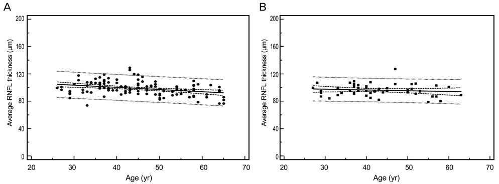
Fig. 1 Relationship between age and average peripapillary retinal nerve fiber layer (RNFL) thickness in long (A) and short-to-normal length (B) eyes.
Cited by 1 articles
-
Retinal Nerve Fiber Layer and Macular Retinal Ganglion Cell Layer Thicknesses in Healthy Korean Children
Yeji Kim, Young Hoon Hwang
J Korean Ophthalmol Soc. 2019;60(9):874-880. doi: 10.3341/jkos.2019.60.9.874.
Reference
-
1. Quigley HA, Dunkelberger GR, Green WR. Chronic human glaucoma causing selectively greater loss of large optic nerve fibers. Ophthalmology. 1988. 95:357–363.2. Airaksinen PJ, Alanko HI. Effect of retinal nerve fibre loss on the optic nerve head configuration in early glaucoma. Graefes Arch Clin Exp Ophthalmol. 1983. 220:193–196.3. Balazsi AG, Rootman J, Drance SM, et al. The effect of age on the nerve fiber population of the human optic nerve. Am J Ophthalmol. 1984. 97:760–766.4. Budenz DL, Anderson DR, Varma R, et al. Determinants of normal retinal nerve fiber layer thickness measured by Stratus OCT. Ophthalmology. 2007. 114:1046–1052.5. Vernon SA, Rotchford AP, Negi A, et al. Peripapillary retinal nerve fibre layer thickness in highly myopic Caucasians as measured by Stratus optical coherence tomography. Br J Ophthalmol. 2008. 92:1076–1080.6. Choi SW, Lee SJ. Thickness changes in the fovea and peripapillary retinal nerve fiber layer depend on the degree of myopia. Korean J Ophthalmol. 2006. 20:215–219.7. Kim JW, Kim YY. Changes in RNFL thickness according to the myopia in patients with glaucoma and ocular hypertension. J Korean Ophthalmol Soc. 2008. 49:1634–1640.8. Ha SW, Rho SH. Age-related differences of optical coherence tomography data in Koreans. J Korean Ophthalmol Soc. 2005. 46:2037–2044.9. Budenz DL, Fredette MJ, Feuer WJ, Anderson DR. Reproducibility of peripapillary retinal nerve fiber thickness measurements with stratus OCT in glaucomatous eyes. Ophthalmology. 2008. 115:661–666.e4.10. Hoffer KJ. Steinert RF, editor. Intraocular lens implant power calculation, selection, and ocular biometry. Cataract surgery: techniques, complications, and management. 2004. 2nd ed. Philadelphia: Elsevier Science;37–38.11. Parikh RS, Parikh SR, Sekhar GC, et al. Normal age-related decay of retinal nerve fiber layer thickness. Ophthalmology. 2007. 114:921–926.12. Sung KR, Wollstein G, Bilonick RA, et al. Effects of age on optical coherence tomography measurements of healthy retinal nerve fiber layer, macula, and optic nerve head. Ophthalmology. 2009. 116:1119–1124.13. Kim MJ, Lee EJ, Kim TW. Peripapillary retinal nerve fibre layer thickness profile in subjects with myopia measured using the Stratus optical coherence tomography. Br J Ophthalmol. 2010. 94:115–120.14. Rauscher FM, Sekhon N, Feuer WJ, Budenz DL. Myopia affects retinal nerve fiber layer measurements as determined by optical coherence tomography. J Glaucoma. 2009. 18:501–505.15. Leung CK, Mohamed S, Leung KS, et al. Retinal nerve fiber layer measurements in myopia: an optical coherence tomography study. Invest Ophthalmol Vis Sci. 2006. 47:5171–5176.
- Full Text Links
-
- Actions
-
Cited
- CITED
-
- Close
- Share
-
- Similar articles
-
- Associations of Peripapillary Retinal Nerve Fiber Layer and Macular Retinal Layer Thickness with Serum Homocysteine Concentration
- Reproducibility of Retinal Nerve Fiber Layer Thickness Evaluation by Nerve Fiber Analyzer
- A Case of Retinal Herniation through Peripapillary Pit Resulting in Retinal Nerve Fiber Layer Defect
- Biometry of Retinal Nerve Fiber Layer Thickness by NFA
- Hierarchical Cluster Analysis of Peripapillary Retinal Nerve Fiber Layer Damage and Macular Ganglion Cell Loss in Open Angle Glaucoma
