Computed Tomography and Magnetic Resonance Imaging of Myoepitheliloma in the Soft Palate: A Case Report
- Affiliations
-
- 1Department of Radiology, Eulji University Hospital, Daejeon, Korea. midosyu@eulji.ac.kr
- 2Department of Pathology, Eulji University Hospital, Daejeon, Korea.
- 3Department of Otorhinolaryngology, Eulji University Hospital, Daejeon, Korea.
- KMID: 1443489
- DOI: http://doi.org/10.3348/jksr.2011.65.1.23
Abstract
- We report the appearance of myoepithelioma arising from minor salivary glands in the soft palate observed on computed tomography (CT) and magnetic resonance imaging (MRI). CT, the tumor was round with a smooth and partial lobulating contour, and slightly marginal contrast enhancement. On T1-weighted images, the mass had heterogeneous iso-signal intensity compared to the pharyngeal muscle. Additionally, the tumor had heterogeneously high T2 signal intensity with heterogeneously strong enhancement on the Gd-enhanced T1-weighted image. Radiologists should consider myoepithelioma in the radiological differential diagnosis of soft palate tumors.
MeSH Terms
Figure
-
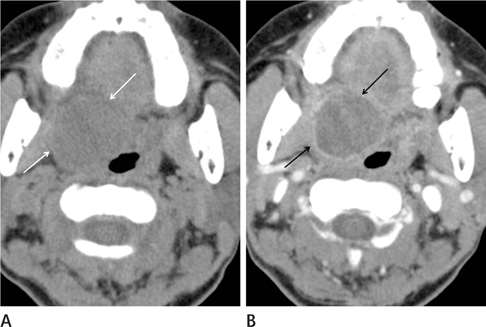
Fig. 1 A 53-year-old woman with a palpable mass on the soft palate. A. Pre-enhanced CT images show a mass on the right soft palate (white arrows). B. On the contrast-enhanced CT images, the mass is faintly enhanced, and shows peripheral wall enhancement (black arrows), suggesting a capsular structure.
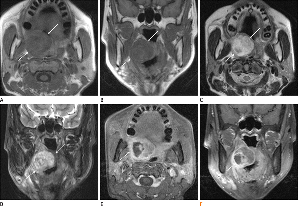
Fig. 2 Axial (A) and coronal (B) T1-weighted images show a slightly heterogeneous mass with iso-signal intensity on the right soft palate (arrows). Axial (C) and coronal (D) T2-weighted images show the heterogeneous hyperintense mass on the right soft palate (arrows). Axial (E) and coronal (F) Gd-enhanced T1-weighted images show a heterogeneously enhancing tumor with a poorly enhancing intratumoral area (arrow), suggesting the presence of a focal necrotic component. A poorly enhancing portion on Gd-enhanced T1-weighted images is nearly consistent with a subtle low attenuated portion on the contrast-enhanced CT images (Fig. 1B).
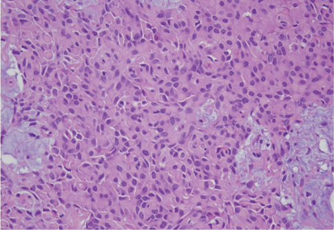
Fig. 3 Haematoxylin and Eosin-stained sections (400 × magnification) of the resected mass show marked plasmacytoid cell proliferation, polygonal cells with eccentric nuclei, and abundant hyaline eosinophilic cytoplasm with some myxoid stroma background. No ductal differentiation is observed. Histologic findings suggest myoepithelioma.
Reference
-
1. Kim HS, Lee WM, Choi SM. Myoepitheliomas of the soft palate: helical CT findings in two patients. Korean J Radiol. 2007; 8:552–555.2. Wang S, Shi H, Wang L, Yu Q. Myoepithelioma of the parotid gland: CT imaging findings. AJNR Am J Neuroradiol. 2008; 29:1372–1375.3. Katsuyama E, Kaneoka A, Higuchi K, Takasu K. Myoepithelioma of the soft palate. Acta Cytol. 1997; 41:1856–1858.4. Seifert G, Brocheriou C, Cardesa A, Eveson JW. WHO International Histological Classification of Tumours. Tentative Histological Classification of Salivary Gland Tumours. Pathol Res Pract. 1990; 186:555–581.5. Monzen Y, Fukushima N, Fukuhara T. Myoepithelioma and malignant myoepithelioma arising from the salivary gland: computed tomography and magnetic resonance findings. Australas Radiol. 2007; 51:B169–B172.6. Hiwatashi A, Matsumoto S, Kamoi I, Yamashita H, Nakashima A. Imaging features of myoepithelioma arising from the hard palate. A case report. Acta Radiol. 2000; 41:417–419.7. Suba Z, Németh Z, Gyulai-Gaál S, Ujpál M, Szende B, Szabó G. Malignant myoepithelioma. Clinicopathological and immunohistochemical characteristics. Int J Oral Maxillofac Surg. 2003; 32:339–341.8. Savera AT, Sloman A, Huvos AG, Klimstra DS. Myoepithelial carcinoma of the salivary glands: a clinicopathologic study of 25 patients. Am J Surg Pathol. 2000; 24:761–774.9. Pons Vicente O, Almendros Marqués N, Berini Aytés L, Gay Escoda C. Minor salivary gland tumors: a clinicopathological study of 18 cases. Med Oral Patol Oral Cir Bucal. 2008; 13:E582–E588.
