The T2-Shortening Effect of Gadolinium and the Optimal Conditions for Maximizing the CNR for Evaluating the Biliary System: a Phantom Study
- Affiliations
-
- 1Department of Radiology and the Research Institute of Radiological Science, Severance Children's Hospital, Yonsei University, College of Medicine, Seoul 120-752, Korea. mjkim@yuhs.ac
- 2Department of Radiology, Gangnam Severance Hospital, Yonsei University, College of Medicine, Seoul 135- 720, Korea.
- 3Department of Internal Medicine, Severance Hospital, Yonsei University, College of Medicine, Seoul 120-752, Korea.
- 4Department of Pharmacology, Yonsei University, College of Medicine, Seoul 120-752, Korea.
- 5Department of Radiology, Seoul National University, College of Medicine, Seoul National University Hospital, Seoul 110-744, Korea.
- KMID: 1122333
- DOI: http://doi.org/10.3348/kjr.2011.12.3.358
Abstract
OBJECTIVE
Clear depiction of the common bile duct is important when evaluating neonatal cholestasis in order to differentiate biliary atresia from other diseases. During MR cholangiopancreatography, the T2-shortening effect of gadolinium can increase the contrast-to-noise ratio (CNR) of the bile duct and enhance its depiction. The purpose of this study was to confirm, by performing a phantom study, the T2-shortening effect of gadolinium, to evaluate the effect of different gadolinium chelates with different gadolinium concentrations and different magnetic field strengths for investigating the optimal combination of these conditions, and for identifying the maximum CNR for the evaluation of the biliary system.
MATERIALS AND METHODS
MR imaging using a T2-weighted single-shot fast spin echo sequence and T2 relaxometry was performed with a sponge phantom in a syringe tube. Two kinds of contrast agents (Gd-DTPA and Gd-EOB-DTPA) with different gadolinium concentrations were evaluated with 1.5T and 3T scanners. The signal intensities, the CNRs and the T2 relaxation time were analyzed.
RESULTS
The signal intensities significantly decreased as the gadolinium concentrations increased (p < 0.001) with both contrast agents. These signal intensities were higher on a 3T (p < 0.001) scanner. The CNRs were higher on a 1.5T (p < 0.001) scanner and they showed no significant change with different gadolinium concentrations. The T2 relaxation time also showed a negative correlation with the gadolinium concentrations (p < 0.001) and the CNRs showed decrease more with Gd-EOB-DTPA (versus Gd-DTPA; p < 0.001) on a 3T scanner (versus 1.5T; p < 0.001).
CONCLUSION
A T2-shortening effect of gadolinium exhibits a negative correlation with the gadolinium concentration for both the signal intensities and the T2 relaxation time. A higher CNR can be obtained with Gd-DTPA on a 1.5T MRI scanner.
MeSH Terms
-
Analysis of Variance
Biliary Atresia/diagnosis
Cholangiopancreatography, Magnetic Resonance/*methods
Cholestasis/diagnosis
Contrast Media/*administration & dosage/diagnostic use
Diagnosis, Differential
Gadolinium DTPA/*administration & dosage/diagnostic use
Humans
Image Processing, Computer-Assisted
Linear Models
*Phantoms, Imaging
Figure
-
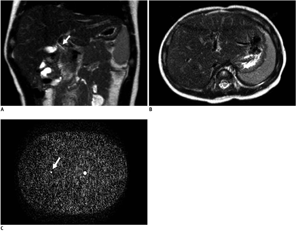
Fig. 1 Size comparison between normal neonate liver, common bile duct and sponge phantom. T2-weighted coronal (A) and axial (B) images of normal neonate liver and axial image of sponge phantom (C) (all magnified by same factor) seem to be similar in size. Also, diameter of common bile duct (2 mm, arrow in A) in normal neonate is similar with inner diameter of 1 cc syringe tube (3 mm, arrow in C) in sponge phantom.
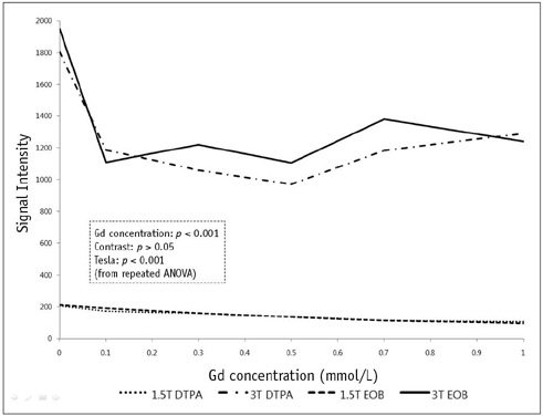
Fig. 2 Line chart depicting relationship between signal intensities and gadolinium concentrations with different gadolinium chelates and different magnetic field strengths. Signal intensities of sponge phantom decreased as concentration of gadolinium chelate increased under all conditions; these were higher on 3T. ANOVA = analysis of variance, DTPA = Gd-DTPA, EOB = Gd-EOB-DTPA
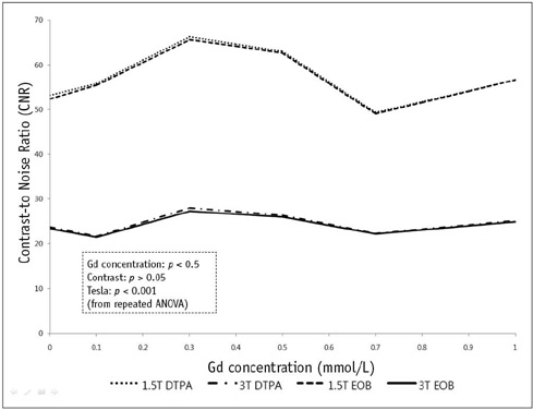
Fig. 3 Line chart depicting relationship between contrastto-noise ratios and gadolinium concentrations with different gadolinium chelates on different magnetic field strengths. Contrast-to-noise radios of tube in sponge phantom were not correlated with gadolinium concentration. However, contrast-to-noise ratios were higher on 1.5T with both contrast agents. ANOVA = analysis of variance, DTPA = Gd-DTPA, EOB = Gd-EOB-DTPA
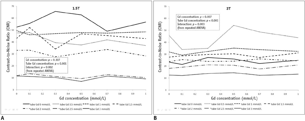
Fig. 4 Line chart depicting relationship between contrast-to-noise ratios and gadolinium concentrations with Gd-EOB-DTPA on 1.5T (A) and 3T (B). Different tube gadolinium concentrations (on both magnetic field strengths) and sponge gadolinium concentration changes (on 3T) had significantly influence on the contrast-to-noise ratios. ANOVA = analysis of variance
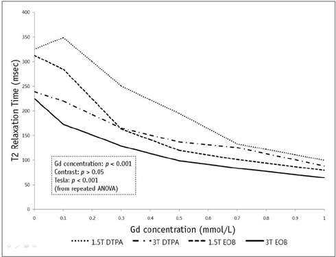
Fig. 5 Line chart depicting relationship between T2 relaxation time and gadolinium concentration with different gadolinium chelates on different magnetic field strengths. T2 relaxation time decreased as gadolinium concentration increased in all conditions. T2 relaxation times were longer with Gd-DTPA on 1.5T. ANOVA = analysis of variance, DTPA = Gd-DTPA, EOB = Gd-EOB-DTPA
Cited by 1 articles
-
Evaluation of the Subscapularis Tendon Tears on 3T Magnetic Resonance Arthrography: Comparison of Diagnostic Performance of T1-Weighted Spectral Presaturation with Inversion-Recovery and T2-Weighted Turbo Spin-Echo Sequences
Hoseok Lee, Joong Mo Ahn, Yusuhn Kang, Joo Han Oh, Eugene Lee, Joon Woo Lee, Heung Sik Kang
Korean J Radiol. 2018;19(2):320-327. doi: 10.3348/kjr.2018.19.2.320.
Reference
-
1. Carlos RC, Hussain HK, Song JH, Francis IR. Gadolinium-ethoxybenzyl-diethylenetriamine pentaacetic acid as an intrabiliary contrast agent: preliminary assessment. AJR Am J Roentgenol. 2002. 179:87–92.2. Takaya J, Nakano S, Imai Y, Fujii Y, Kaneko K. Usefulness of magnetic resonance cholangiopancreatography in biliary structures in infants: a four-case report. Eur J Pediatr. 2007. 166:211–214.3. Jaw TS, Kuo YT, Liu GC, Chen SH, Wang CK. MR cholangiography in the evaluation of neonatal cholestasis. Radiology. 1999. 212:249–256.4. Rohrer M, Bauer H, Mintorovitch J, Requardt M, Weinmann HJ. Comparison of magnetic properties of MRI contrast media solutions at different magnetic field strengths. Invest Radiol. 2005. 40:715–724.5. Kanematsu M, Matsuo M, Shiratori Y, Kondo H, Hoshi H, Yasuda I, et al. Thick-section half-Fourier rapid acquisition with relaxation enhancement MR cholangiopancreatography: effects of i.v. administration of gadolinium chelate. AJR Am J Roentgenol. 2002. 178:755–761.6. Kuperman VY, Alley MT. Differentiation between the effects of T1 and T2* shortening in contrast-enhanced MRI of the breast. J Magn Reson Imaging. 1999. 9:172–117.7. Elster AD, Sobol WT, Hinson WH. Pseudolayering of Gd-DTPA in the urinary bladder. Radiology. 1990. 174:379–381.8. May DA, Pennington DJ. Effect of gadolinium concentration on renal signal intensity: an in vitro study with a saline bag model. Radiology. 2000. 216:232–236.9. Hwang HS, Kim SH, Jeon TY, Choi D, Lee WJ, Lim HK. Hypointense hepatic lesions depicted on gadobenate dimeglumine-enhanced three-hour delayed hepatobiliary-phase MR imaging: differentiation between benignancy and malignancy. Korean J Radiol. 2009. 10:294–302.10. Bollow M, Taupitz M, Hamm B, Staks T, Wolf KJ, Weinmann HJ. Gadolinium-ethoxybenzyl-DTPA as a hepatobiliary contrast agent for use in MR cholangiography: results of an in vivo phase-I clinical evaluation. Eur Radiol. 1997. 7:126–132.11. Strich G, Hagan PL, Gerber KH, Slutsky RA. Tissue distribution and magnetic resonance spin lattice relaxation effects of gadolinium-DTPA. Radiology. 1985. 154:723–726.12. Weinmann HJ, Brasch RC, Press WR, Wesbey GE. Characteristics of gadolinium-DTPA complex: a potential NMR contrast agent. AJR Am J Roentgenol. 1984. 142:619–624.13. Bellin MF, Webb JA, Van Der Molen AJ, Thomsen HS, Morcos SK. Safety of MR liver specific contrast media. Eur Radiol. 2005. 15:1607–1614.14. Hamm B, Staks T, Muhler A, Bollow M, Taupitz M, Frenzel T, et al. Phase I clinical evaluation of Gd-EOB-DTPA as a hepatobiliary MR contrast agent: safety, pharmacokinetics, and MR imaging. Radiology. 1995. 195:785–792.15. Nakamura Y, Ohmoto T, Saito T, Kajima T, Nishimaru E, Ito K. Effects of gadolinium-ethoxybenzyl-diethylenetriamine pentaacetic acid on T2-weighted MRCP. Magn Reson Med Sci. 2009. 8:143–148.
- Full Text Links
-
- Actions
-
Cited
- CITED
-
- Close
- Share
-
- Similar articles
-
- Flow Signal Characteristics in 3 Dimensional Time of Flight MR Angiography Using Flow Phantom
- Usefulness of Postcontrast T2-Weighted Images in Shortening the Total Scan Time of a Gadoxectic Acid Enhanced MRI of the Liver: a Comparison between Precontrast and Postcontrast T2-Weighted Images
- Double Contrast Media enhanced MRI with Ferumoxides-Gadolinium on Hepatocellular Carcinoma
- The Production and Evaluation of the Tissue-equivalent Phantom for the Magnetic Resonance Imaging
- Optimizations of 3D MRI Techniques in Brain by Evaluating SENSE Factors
