Clin Orthop Surg.
2010 Sep;2(3):167-172. 10.4055/cios.2010.2.3.167.
Evaluation of Shoulder Disorders by 2-[F-18]-fluoro-2-deoxy-D-glucose Positron Emission Tomography and Computed Tomography
- Affiliations
-
- 1Department of Orthopaedic Surgery, Chosun University College of Medicine, Gwangju, Korea. orthoped@chosun.ac.kr
- KMID: 999436
- DOI: http://doi.org/10.4055/cios.2010.2.3.167
Abstract
- BACKGROUND
Although flourine-18-flourodeoxyglucose (FDG) positron emission tomography (PET) has a limitation for localizing anatomical structures, combining it with computed tomography (CT) has made it more efficient for overcoming such limitations. This study aims to evaluate the efficacy of PET/CT for evaluating diseases of the shoulder.
METHODS
Retrospective examination was performed on 25 patients who underwent FDG-PET/CT scanning. All the patients were over 60 years of age, and they were evaluated both clinically and radiologically for shoulder pain. The study period was from May, 2006 to May, 2008. One of the patients had metastatic lesion in a shoulder and this patient was excluded from the study, so the total number of subjects in the study was finally 24 patients.
RESULTS
PET/CT showed 67% sensitivity, 73% specificity, a positive predictive value of 60%, a negative predictive value of 79%, 27% false positivity and 33% false negativity concerning shoulder pain. PET/CT showed negative finding in 4 cases that were successfully treated by operative treatment (rotator cuff tear [RCT], 3 cases; impingement syndrome, 1 case). Negative findings were also noted in 6 cases in which the pain subsided after conservative treatment (RCT, 1 case; suspected RCT, 2 cases; impingement syndrome, 3 cases). All the patients with osteoarthritis and rheumatoid arthritis had positive findings on PET/CT scanning.
CONCLUSIONS
PET/CT is a useful adjunct to the existing imaging modalities to assess functional and pathophysiologic processes and at a very early stage, and so PET/CT can help physicians make better preoperative and postoperative decisions on treatment.
MeSH Terms
-
Aged
Bursitis/radiography/radionuclide imaging
Female
Fluorodeoxyglucose F18/*diagnostic use
Humans
Joint Diseases/*radiography/*radionuclide imaging
Male
Middle Aged
*Positron-Emission Tomography
Predictive Value of Tests
Radiopharmaceuticals/*diagnostic use
Rotator Cuff/injuries/radiography/radionuclide imaging
Sensitivity and Specificity
Shoulder Joint/*radiography/*radionuclide imaging
*Tomography, X-Ray Computed
Figure
-

Fig. 1 Magnetic resonance imaging of a 66-year-old male with a symptomatic rotator cuff tear (A) and the postoperative radiograph after the repair (B). Fourteen month after the cuff repair, the positron emission tomography / computed tomography revealed normal flourine-18-flourodeoxyglucose uptake (C), signifying relief from the previous symptoms.
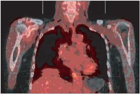
Fig. 2 A 69-year-old female patient with rheumatoid arthritis in the shoulder shows generalized increased uptake on the left side.
Reference
-
1. Nehmeh SA, Erdi YE, Ling CC, et al. Effect of respiratory gating on reducing lung motion artifacts in PET imaging of lung cancer. Med Phys. 2002. 29(3):366–371.
Article2. Bunker T. Rotator cuff disease. Curr Orthop. 2002. 16(3):223–233.
Article3. Vlychou M, Dailiana Z, Fotiadou A, Papanagiotou M, Fezoulidis IV, Malizos K. Symptomatic partial rotator cuff tears: diagnostic performance of ultrasound and magnetic resonance imaging with surgical correlation. Acta Radiol. 2009. 50(1):101–105.
Article4. Al-Shawi A, Badge R, Bunker T. The detection of full thickness rotator cuff tears using ultrasound. J Bone Joint Surg Br. 2008. 90(7):889–892.
Article5. Meyers PR, Craig JG, van Holsbeeck M. Shoulder ultrasound. AJR Am J Roentgenol. 2009. 193(3):W174.
Article6. Teefey SA, Petersen B, Prather H. Shoulder Ultrasound vs MRI for rotator cuff pathology. PM R. 2009. 1(5):490–495.
Article7. Allen GM. Shoulder ultrasound imaging-integrating anatomy, biomechanics and disease processes. Eur J Radiol. 2008. 68(1):137–146.
Article8. Kolla S, Motamedi K. Ultrasound evaluation of the shoulder. Semin Musculoskelet Radiol. 2007. 11(2):117–125.
Article9. Daenen B, Houben G, Bauduin E, Lu KV, Meulemans JL. Ultrasound of the shoulder. JBR-BTR. 2007. 90(5):325–337.10. Beggs I. Ultrasound of the shoulder and elbow. Orthop Clin North Am. 2006. 37(3):277–285. v
Article11. Moosmayer S, Smith HJ. Diagnostic ultrasound of the shoulder: a method for experts only? Results from an orthopedic surgeon with relative inexpensive compared to operative findings. Acta Orthop. 2005. 76(4):503–508.12. Lick-Schiffer W. Ultrasound examination of the shoulder joint. Wien Med Wochenschr. 1996. 146(6-7):121–123.13. Gustav K. Clinical molecular anatomic imaging. 2003. Philadelphia: Lippincott Williams & Wilkins;251–270. 412–428.14. Wandler E, Kramer EL, Sherman O, Babb J, Scarola J, Rafii M. Diffuse FDG shoulder uptake on PET is associated with clinical findings of osteoarthritis. AJR Am J Roentgenol. 2005. 185(3):797–803.
Article15. Suzuki H, Watanabe H, Shinozaki T, Yanagawa T, Suzuki R, Takagishi K. Positron emission tomography imaging of musculoskeletal tumors in the shoulder girdle. J Shoulder Elbow Surg. 2004. 13(6):635–647.
Article16. Shinozaki T, Takagishi K, Ohsawa T, Yamaji T, Endo K. Pre- and postoperative evaluation of the metabolic activity in muscles associated with ruptured rotator cuffs by F-FDG PET imaging. Clin Physiol Funct Imaging. 2006. 26(6):338–342.
Article17. Kim CH, Kim SH, Hyun OJ, et al. Usefulness of 18F-FDG-PET/CT in the diagnosis of cervical lymph node metastases of head and neck cancer. Korean J Nucl Med. 2005. 39(5):269–277.18. Chung JK. Mechanisms of glucose uptake in cancer tissue. Korean J Nucl Med. 1999. 33(1):1–10.19. Schmitz A, Risse JH, Grunwald F, Gassel F, Biersack HJ, Schmitt O. Fluorine-18 fluorodeoxyglucose positron emission tomography findings in spondylodiscitis: preliminary results. Eur Spine J. 2001. 10(6):534–539.
Article20. Shinozaki T, Takagishi K, Ichikawa A, et al. Use of 2-[18F]-fluoro-2-deoxy-d-glucose positron emission tomography (FDG PET) imaging for the evaluation of muscle metabolic activity in ruptured rotator cuffs: identification of shoulder muscles by fusion imaging studies involving both FDG PET and magnetic resonance imaging. J Shoulder Elbow Surg. 2003. 12(6):544–549.
Article21. Ghanem N, Uhl M, Brink I, et al. Diagnostic value of MRI in comparison to scintigraphy, PET, MS-CT and PET/CT for the detection of metastases of bone. Eur J Radiol. 2005. 55(1):41–55.
Article22. Metser U, Lerman H, Blank A, Lievshitz G, Bokstein F, Even-Sapir E. Malignant involvement of the spine: assessment by 18F-FDG PET/CT. J Nucl Med. 2004. 45(2):279–284.23. Gratz S, Dorner J, Fischer U, et al. 18F-FDG hybrid PET in patients with suspected spondylitis. Eur J Nucl Med Mol Imaging. 2002. 29(4):516–524.
Article24. Yamada S, Kubota K, Kubota R, Ido T, Tamahashi N. High accumulation of fluorine-18-fluorodeoxyglucose in turpentine-induced inflammatory tissue. J Nucl Med. 1995. 36(7):1301–1306.25. Lehman C, Cuomo F, Kummer FJ, Zuckerman JD. The incidence of full thickness rotator cuff tears in a large cadaveric population. Bull Hosp Jt Dis. 1995. 54(1):30–31.26. Sher JS, Uribe JW, Posada A, Murphy BJ, Zlatkin MB. Abnormal findings on magnetic resonance images of asymptomatic shoulders. J Bone Joint Surg Am. 1995. 77(1):10–15.
Article27. Tempelhof S, Rupp S, Seil R. Age-related prevalence of rotator cuff tears in asymptomatic shoulders. J Shoulder Elbow Surg. 1999. 8(4):296–299.
Article
- Full Text Links
-
- Actions
-
Cited
- CITED
-
- Close
- Share
-
- Similar articles
-
- 18F-2-Deoxy-2-Fluoro-D-Glucose Positron Emission Tomography: Computed Tomography for Preoperative Staging in Gastric Cancer Patients
- Differential Diagnosis of Adrenal Mass Using Imaging Modality: Special Emphasis on F-18 Fluoro-2-Deoxy-D-Glucose Positron Emission Tomography/Computed Tomography
- Positron Emission Tomography: Application in Pediatric Epilepsy
- Metabolic Activities of Benign Musculoskeletal Tumors Using 2-[ F - 18 ] - Fluoro -2 - deoxy - D - glucose ( FDG ) Positron Emission Tomogrphy ( PET ) ( preliminary report )
- 18F-fluoro-2-deoxy-D-glucose positron emission tomography findings of neurolymphomatosis
