The Effect of Yellow Tinted Intraocular Lenses on the Result of Frequency Doubling Perimetry after Cataract Surgery
- Affiliations
-
- 1Department of Ophthalmology and Visual Science, The Catholic University of Korea College of Medicine, Seoul, Korea. ckjoo@catholic.ac.kr
- KMID: 994408
- DOI: http://doi.org/10.3341/kjo.2011.25.1.29
Abstract
- PURPOSE
To investigate the effect of yellow tinted intraocular lenses (IOLs), implanted in the bag after phacoemulsification, on the result of frequency doubling technique (FDT) perimetry.
METHODS
For 68 eyes of 52 patients, an IOL was implanted in the bag after phacoemulsification. The patients were categorized into three groups according to the type of implanted IOLs used. IOLs were selected randomly among two types of yellow IOLs (Acrysof SN60WF IOL, Hoya YA60BBR IOL) and a clear IOL was used as a control. A FDT Humphrey matrix was performed 2 months after the surgery. The mean deviation (MD) and pattern standard deviation (PSD) among these three groups was analyzed using Mann-Whitney U-test.
RESULTS
Two months after the procedure, there was no significant difference between each of the three groups: the clear IOL and Hoya YA60BBR IOL (MD, p = 0.21; PSD, p = 0.27), the clear IOL and Alcon SN60WF IOL (MD, p = 0.11; PSD, p = 0.22), and the Hoya YA60BBR IOL and Alcon SN60WF IOL (MD, p = 0.33; PSD, p = 0.56).
CONCLUSIONS
When interpreting the result of the FDT after cataract surgery, the color and type of IOLs used should not be considered.
MeSH Terms
Figure
-

Fig. 1 Postoperative mean deviation (MD) and pattern standard deviation (PSD) of the frequency doubling perimetry result. There was no significant difference in MD between the clear and Hoya YA60BBR intraocular lens (IOL) (Mann-Whitney U-test, p = 0.21). There was no significant difference in PSD between clear and Hoya YA60BBR IOLs (Mann-Whitney U-test, p = 0.27).

Fig. 2 Postoperative mean deviation (MD) and pattern standard deviation (PSD) of the frequency doubling perimetry result. There was no significant difference in MD between the clear and Alcon SN60WF intraocular lenses (IOLs) (Mann-Whitney U-test, p = 0.11). There was no significant difference in PSD between the clear and Alcon SN60WF IOLs (Mann-Whitney U-test, p = 0.22).
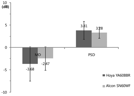
Fig. 3 Postoperative mean deviation (MD) and pattern standard deviation (PSD) of the frequency doubling perimetry result. There was no significant difference in MD between the Hoya YA60BBR and Alcon SN60WF intraocular lenses (IOLs) (Mann-Whitney U-test, p = 0.33). There was no significant difference in PSD between the Hoya YA60BBR and Alcon SN60WF IOLs (Mann-Whitney U-test, p = 0.56).
Reference
-
1. Yanagi Y, Inoue Y, Iriyama A, Jang WD. Effects of yellow intraocular lenses on light-induced upregulation of vascular endothelial growth factor. J Cataract Refract Surg. 2006. 32:1540–1544.2. Fogagnolo P, Rossetti L, Ranno S, et al. Short-wavelength automated perimetry and frequency-doubling technology perimetry in glaucoma. Prog Brain Res. 2008. 173:101–124.3. Tanna AP, Abraham C, Lai J, Shen J. Impact of cataract on the results of frequency-doubling technology perimetry. Ophthalmology. 2004. 111:1504–1507.4. Kook MS, Yang SJ, Kim S, et al. Effect of cataract extraction on frequency doubling technology perimetry. Am J Ophthalmol. 2004. 138:85–90.5. Ueda T, Ota T, Yukawa E, Hara Y. Frequency doubling technology perimetry after clear and yellow intraocular lens implantation. Am J Ophthalmol. 2006. 142:856–858.6. Ernest PH. Light-transmission-spectrum comparison of foldable intraocular lenses. J Cataract Refract Surg. 2004. 30:1755–1758.7. Brockmann C, Schulz M, Laube T. Transmittance characteristics of ultraviolet and blue-light-filtering intraocular lenses. J Cataract Refract Surg. 2008. 34:1161–1166.8. Lee BB, Smith VC, Pokorny J, Kremers J. Rod inputs to macaque ganglion cells. Vision Res. 1997. 37:2813–2828.9. Purpura K, Kaplan E, Shapley RM. Background light and the contrast gain of primate P and M retinal ganglion cells. Proc Natl Acad Sci U S A. 1988. 85:4534–4537.10. Sun H, Pokorny J, Smith VC. Rod-cone interactions assessed in inferred magnocellular and parvocellular postreceptoral pathways. J Vis. 2001. 1:42–54.11. Stabell U, Stabell B. Mechanisms of chromatic rod vision in scotopic illumination. Vision Res. 1994. 34:1019–1027.12. Sterling P, Freed MA, Smith RG. Architecture of rod and cone circuits to the on-beta ganglion cell. J Neurosci. 1988. 8:623–642.
- Full Text Links
-
- Actions
-
Cited
- CITED
-
- Close
- Share
-
- Similar articles
-
- Contrast Sensitivity and Color Vision Comparison Between Clear and Yellow-Tinted Intraocular Lens in Diabetic Retinopathy
- Effect of Intraocular Lens Implantation on Electro retinogram
- The Effects of Cheap Tinted Contact Lenses on Corneal Swelling and Ocular Surface Inflammation
- Intraindividual Comparison of Visual Outcomes between Blue Light-filtering and Ultraviolet Light-filtering Intraocular Lens
- Implantation of Black Diaphragm Intraocular Lens in Cataract Surgery with Congenital Aniridia
