MRI Appearance of Florid Cystic Endosalpingiosis of the Uterus: a Case Report
- Affiliations
-
- 1Department of MRI, Rajiv Gandhi Cancer Institute & Research Centre, India. s_taneja1974@yahoo.com
- 2Department of Histo-Pathology, Rajiv Gandhi Cancer Institute & Research Centre, India.
- 3Department of Gynaecology and Oncology, Rajiv Gandhi Cancer Institute & Research Centre, India.
- 4Department of Pathology, Rajiv Gandhi Cancer Institute & Research Centre, India.
- 5Department of MRI, Rajiv Gandhi Cancer Institute & Research Centre, India.
- KMID: 984903
- DOI: http://doi.org/10.3348/kjr.2010.11.4.476
Abstract
- Endosalpingiosis is a non-neoplastic proliferation of ectopic tubal epithelium. It may be found incidentally or the patients may present with chronic pelvic pain. It may resemble a gynecologic malignancy on imaging findings and clinicians and radiologists should be aware of this benign entity to render a correct diagnosis and to avoid over-treatment. We report here the MR imaging appearance of a case of florid cystic endosalpingiosis.
MeSH Terms
Figure
-
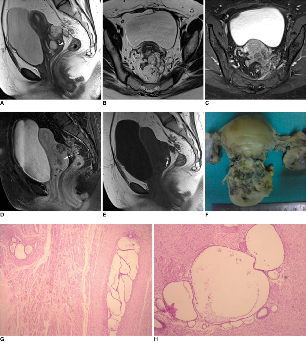
Fig. 1 Florid cystic endosalpingiosis of uterus in 40-year-old woman. A-C. Sagittal T2-weighted (A), axial T2-weighted (B) and axial post-contrast fat suppressed T1-weighted (C) images show heterogeneously enhancing complex cystic mass involving posterior myometrium at uterocervical junction and extending into pouch of Douglas. Discrete cystic lesion is seen in posterior cervical stroma and this appears as being predominantly hyperintense on T2-weighted images (arrow). D, E. Sagittal post-contrast fat suppressed T1-weighted image (D) shows no significant post-contrast enhancement in discrete cystic lesion in posterior cervical stroma (arrow). Sagittal pre-contrast T1-weighted image (E) shows small areas of hyperintensity interspersed within cystic lesion (arrow). There was no evidence of hemorrhage within lesion on pathologic examination and small areas of hyperintensity were presumed to be mucinous content. F. Resected specimen of corpus uteri and cervix shows irregular grayish white tissue adherent to posterior serosal surface. G, H. Microphotograph showing multiple foci of cystically dilated glands in posterior cervical stroma (Hematoxylin & Eosin stain × 100) (G). Microphotograph showing circular gland lined by tubal epithelium. Adjoining cystic structures also noted (H).
Reference
-
1. Clement PB, Young RH. Florid cystic endosalpingiosis with tumor like manifestations: a report of four cases including the first reported cases of transmural endosalpingiosis of the uterus. Am J Surg Pathol. 1999. 23:166–175.2. Bazot M, Vacher Lavenu MC, Bigot JM. Imaging of endosalpingiosis. Clin Radiol. 1999. 54:482–485.3. Zinsser KR, Wheeler JE. Endosalpingiosis in the omentum: a study of autopsy and surgical material. Am J Surg Pathol. 1982. 6:109–117.4. deHoop TA, Mira J, Thomas MA. Endosalpingiosis and chronic pelvic pain. J Reprod Med. 1997. 42:613–616.5. Cil AP, Atasoy P, Kara SA. Myometrial involvement of tumor-like cystic endosalpingiosis: a rare entity. Ultrasound Obstet Gynecol. 2008. 32:106–110.6. Tutschka BG, Lauchlan SC. Endosalpingiosis. Obstet Gynecol. 1980. 55:S57–S60.7. Young RH, Clement PB. Müllerianosis of the urinary bladder. Mod Pathol. 1996. 9:731–737.8. Cajigas A, Axiotis CA. Endosalpingiosis of the vermiform appendix. Int J Gynaecol Pathol. 1990. 9:291–295.9. Shim SH, Kim HS, Joo M, Chang SH, Kwak JE. Florid cystic endosalpingiosis of the uterus: a case report. Korean J Pathol. 2008. 42:189–191.10. Heatley MK, Russell P. Florid cystic endosalpingiosis of the uterus. J Clin Pathol. 2001. 54:399–400.
- Full Text Links
-
- Actions
-
Cited
- CITED
-
- Close
- Share
-
- Similar articles
-
- Multicentric Florid Cystic Endosalpingiosis in Different Anatomical Spaces: A Case Report
- Florid Cystic Endosalpingiosis of the Uterus: A Case Report
- Sonographic Appearance of Endosalpingiosis
- Endosalpingiosis in Postmenopausal Elderly Women
- CT and MR Imaging Findings of Cystic Adenomyosis of the Uterus: Report of Three Cases
