A Case of Chorioretinal Coloboma in a Patient with Achondroplasia
- Affiliations
-
- 1Department of Ophthalmology, Gyeongsang National University School of Medicine, Jinju, Korea. YJM@nongae.gsnu.ac.kr
- 2Gyeongsang Institute of Health Science, Gyeongsang National University, Jinju, Korea.
- KMID: 974325
- DOI: http://doi.org/10.3341/kjo.2010.24.5.302
Abstract
- Achondroplasia is a congenital disorder resulting from a specific disturbance in endochondral bone formation. The ophthalmic features reportedly associated with achondroplasia are telecanthus, exotropia, inferior oblique overaction, angle anomalies and cone-rod dystrophy. This is first report of chorioretinal coloboma in achondroplasia. An 8-year-old female was diagnosed with a developmental delay, known as achondroplasia, seven months after birth. Upon her initial visit, visual acuity was 0.3 in both eyes. The patient had telecanthus but normal ocular motility. Findings were normal upon anterior segment examination. Fundus examination of both eyes revealed about 1,500 microm sized chorioretinal coloboma inferior to the optic nerve head. Upon fluorescent angiography, there was chorioretinal coloboma without any other lesions. Afterward, there was no change in the fundus lesion, and best corrected visual acuity was 0.6 in both eyes. Chorioretinal coloboma is associated with choroidal and retinal detachment. As chorioretinal coloboma and achondroplasia are developmental disorders in the embryonic stage, early detection and regular ophthalmologic examination would be essential in patients with achondroplasia.
Keyword
MeSH Terms
Figure
-

Fig. 1 Clinical photograph of an 8-year-old girl presenting as telecanthus, with pudgy, excessively thick fingers.

Fig. 2 Roentgenogram of the patient. There is caudal narrowing of interpeduncular spaces in the lumbar area (A) and characteristic short limbs with relatively short proximal bone (B,C).
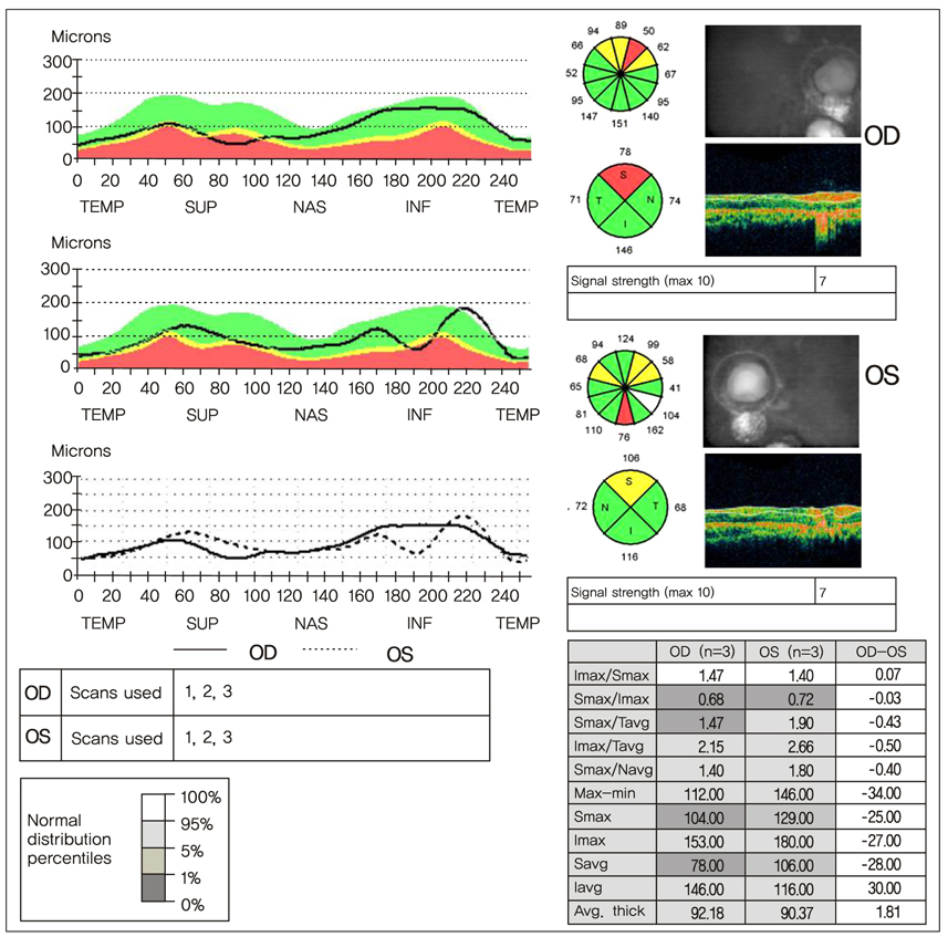
Fig. 3 Optical coherence tomography of the patient shows a superior retinal nerve fiber layer defect in both eyes. TEMP=temporal; SUP=superior; NAS=nasal; INF=inferior; Imax=inferior maximum; Smax=superior maximum; Tavg=temporal average; Navg=nasal average; Savg=superior average; Iavg=inferior average.

Fig. 4 Color photograph of fundus showing chorioretinal coloboma in both eyes.

Fig. 5 Fluorescein angiography of the patient showing only coloboma without other retinal lesions. (A) Early phase. (B) Late phase.
Reference
-
1. Vajo Z, Francomano CA, Wilkin DJ. The molecular and genetic basis of fibroblast growth factor receptor 3 disorders: the achondroplasia family of skeletal dysplasias, Muenke craniosynostosis, and Crouzon syndrome with acanthosis nigricans. Endocr Rev. 2000. 21:23–39.2. Richette P, Bardin T, Stheneur C. Achondroplasia: from genotype to phenotype. Joint Bone Spine. 2008. 75:125–130.3. Maroteaux P. Maroteaux P, editor. Osteochondrodysplasie. Les maladies osseuses de lénfant. 1998. 3rd ed. Paris: Medicine-Sciences Flammarion;55–56.4. Rosenthal AR, Ryan SJ Jr, Horowitz P. Ocular manifestations of dwarfism. Trans Am Acad Ophthalmol Otolaryngol. 1972. 76:1500–1518.5. DeRespinis PA, Caputo AR, Wagner RS, Guo S. Duane's retraction syndrome. Surv Ophthalmol. 1993. 38:257–288.6. Guirgis MF, Thornton SS, Tychsen L, Lueder GT. Cone-rod retinal dystrophy and Duane retraction syndrome in a patient with achondroplasia. J AAPOS. 2002. 6:400–401.7. Garg R, Gupta N, D'Souza P. Fundus albipunctatus in a patient with achondroplasia. J Pediatr Ophthalmol Strabismus. 2007. 44:305–306.8. Barishak YR. Embryology of the eye and its adnexae. Dev Ophthalmol. 1992. 24:1–142.9. Maumenee IH, Mitchell TN. Colobomatous malformations of the eye. Trans Am Ophthalmol Soc. 1990. 88:123–132.10. Daufenbach DR, Ruttum MS, Pulido JS, Keech RV. Chorioretinal colobomas in a pediatric population. Ophthalmology. 1998. 105:1455–1458.
- Full Text Links
-
- Actions
-
Cited
- CITED
-
- Close
- Share
-
- Similar articles
-
- A Case of Rubinstein-Taybi Syndrome with Optic Disc Coloboma and Chorioretinal Coloboma
- Unilateral Peters' Anomaly with Chorioretinal Coloboma in the Other Eye
- A Case of Chorioretinal Coloboma in Triple X Syndrome
- The Development of Recurrent Choroidal Neovascularization in a Patient with Choroidal Coloboma
- Two cases of congenital macular coloboma
