The Relationship between the Visual Prognoses of Branch Retinal Artery Obstruction and Foveal Thickness on OCT
- Affiliations
-
- 1Department of Ophthalmology, Kim's Eye Hospital, Konyang University College of Medicine, Seoul, Korea. -medical-@hanmail.net
- KMID: 974324
- DOI: http://doi.org/10.3341/kjo.2010.24.5.297
Abstract
- PURPOSE
To determine the correlation between the prognosis of branch retinal artery obstruction (BRAO) and the foveal thickness or outer nuclear layer (ONL) thickness on optical coherence tomography (OCT).
METHODS
Twenty-one eyes (21 patients) in patients with resolved, non-complicated BRAO and a normal control of 10 eyes (10 volunteers) were used in this study. The average macular thickness, foveal thickness and ONL thickness at central fovea were measured in both the patients and the control group using spectral domain OCT. The thickness between the patient group and the control group were compared and correlation between the best corrected visual acuity (BCVA) and each thickness was determined.
RESULTS
The average age of the patients was 52 +/- 5.8 years. The average macular thickness, foveal thickness and ONL thickness at the central fovea of the patients were significantly (p < 0.001, p = 0.023, p = 0.021, respectively) thinner than that of the control group. Both the foveal thickness (r(s) = 0.56, p = 0.008) and ONL thickness (r(s) = 0.86, p < 0.001) were significantly correlated with BCVA. There was no significant correlation between the average macular thickness and BCVA.
CONCLUSIONS
The foveal thickness and ONL thickness at the fovea was positively correlated with the BCVA in patients with resolved BRAO.
Keyword
MeSH Terms
Figure
-
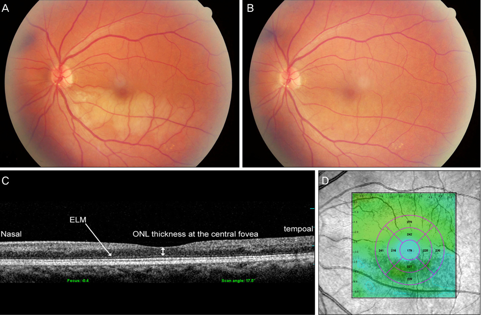
Fig. 1 Color fundus photographs and optical coherence tomography (OCT) image of branch retinal artery obstruction (BRAO) patient (Table 1, No. 2.). (A) Initial color fundus photograph of the left eye of a 43-year-old woman with BRAO whose best corrected visual acuity (BCVA) was 0.1. (B) Color fundus photograph of the same patient after 6 months. Her BCVA was 0.7. (C) Cross-sectional macular image with a 6-mm horizontal line scan across the central fovea. The thickness of outer nuclear laye at the central fovea is approximately 138 µm. (D) Topography image of OCT in the same patient. The average macular thickness is 179 µm, the thickness of the fovea is 221 µm. ELM=external limiting membrane; ONL=outer nuclear layer.
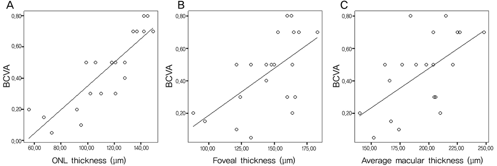
Fig. 2 Graph showing the relationship between best corrected visual acuity (BCVA) of 21 branch retinal artery obstruction eyes and outer nuclear layer (ONL) thickness, foveal thickness, average macular thickness, respectively (Spearman-rank correlation coefficient). (A) The thickness of ONL is positively correlated with BCVA (rs=0.86, p<0.001). (B) The foveal thickness is positively correlated with BCVA (rs=0.56, p=0.008). (C) There is no statically significant correlation between average macular thickness and BCVA (rs=0.41, p=0.186).
Reference
-
1. Brown GC, Shields JA. Cilioretinal arteries and retinal arterial occlusion. Arch Ophthalmol. 1979. 97:84–92.2. Ros MA, Magargal LE, Uram M. Branch retinal-artery obstruction: a review of 201 eyes. Ann Ophthalmol. 1989. 21:103–107.3. Brown GC, Magargal LE, Shields JA, et al. Retinal arterial obstructions in children and young adults. Ophthalmology. 1981. 88:18–25.4. Ryan SJ, Schachat AP, Murphy RP, editors. Retina. Medical retina. 2001. Vol. 2:3rd ed. St. Louis: Mosby;1350–1367.5. Arruga J, Sanders M. Ophthalmologic findings in 70 patients with evidence of retinal embolism. Ophthalmology. 1982. 89:1336–1347.6. Mason JO 3rd, Shah AA, Vail RS, et al. Branch retinal artery occlusion: visual prognosis. Am J Ophthalmol. 2008. 146:455–457.7. Schmidt D, Kube T, Feltgen N. Central retinal artery occlusion: findings in optical coherence tomography and functional correlations. Eur J Med Res. 2006. 11:250–252.8. Hayreh SS, Zimmerman MB, Kimura A, Sanon A. Central retinal artery occlusion. Retinal survival time. Exp Eye Res. 2004. 78:723–736.9. Leung CK, Tham CC, Mohammed S, et al. In vivo measurements of macular and nerve fibre layer thickness in retinal arterial occlusion. Eye (Lond). 2007. 21:1464–1468.10. Asefzadeh B, Ninyo K. Longitudinal analysis of retinal changes after branch retinal artery occlusion using optical coherence tomography. Optometry. 2008. 79:85–89.11. Hayreh SS, Podhajsky PA, Zimmerman MB. Branced retinal arterial occlusion: natural history of visual outcome. Ophthalmology. 2009. 116:1188–1194.12. Nickla DL, Wallman J. The multifunctional choroid. Prog Retin Eye Res. 2010. 29:144–168.13. Matsumoto H, Sato T, Kishi S. Outer nuclear layer thickness at the fovea determines visual outcomes in resolved central serous chorioretinopathy. Am J Ophthalmol. 2009. 148:105–110.
- Full Text Links
-
- Actions
-
Cited
- CITED
-
- Close
- Share
-
- Similar articles
-
- Relationship between Visual Acuity and Photoreceptor Layer or Foveal Thickness on Optical Coherence Tomography after Macular Hole Surgery
- Foveal Retinal Detachment Diagnosed by Optical Coherence Tomography after Successful Retinal Detachment Surgery
- Clinical Results of Vitrectomy in Macular Hemorrhage From a Ruptured Retinal Artery Macroaneurysm
- Optical Coherence Tomography Measurement and Visual Outcome in Acute Central Retinal Artery Occlusion
- The Correlation between Visual Acuity and Patterns of Diabetic Macular Edema in OCT Images
