Anterior Segment Parameters Using Pentacam and Prediction of Corneal Endothelial Cell Loss after Cataract Surgery
- Affiliations
-
- 1Department of Ophthalmology, St. Vincent's Hospital, The Catholic University of Korea School of Medicine, Suwon, Korea.
- 2Department of Ophthalmology, Seoul St. Mary's Hospital, The Catholic University of Korea School of Medicine, Seoul, Korea. mskim@catholic.ac.kr
- KMID: 974322
- DOI: http://doi.org/10.3341/kjo.2010.24.5.284
Abstract
- PURPOSE
We evaluated various preoperative anterior segment parameters measured with a Pentacam rotating Scheimpflug camera and compared them with those of conventional methods. We also evaluated the effect of different parameters on corneal endothelial cells after cataract surgery.
METHODS
Pentacam examination was performed in 88 eyes from 88 patients to evaluate central anterior chamber depth (ACD(pentacam)), nuclear density (Densitometry(pentacam)), anterior chamber volume (ACV), and lens thickness (LT(pentacam)). We compared values of ACD(pentacam) with those of ultrasound (ACD(sono)) and also compared Densitometry(pentacam) values with those of Lens Opacities Classification System (LOCS III) classification. We evaluated the effect of the following preoperative values measured with Pentacam on postoperative endothelial cell loss: pupil size measured both preoperatively and before capsulorrhexsis (Pupil(CCC)), amount of viscoelastics, and LT measured by ultrasound (LT(sono)).
RESULTS
A significant concordance was found between the two grading methods of nuclear opacity: Densitometry(pentacam) and LOCS III classification (tau(b) = 0.414, p = 0.000). We also found a positive correlation between ACD(pentacam) and ACD(sono) (r = 0.823, p = 0.000) and between ACD(pentacam) and ACV (r = 0.650, p = 0.000). There were significant differences between the results of LT(pentacam) and LT(sono). The final regression model identified Densitometry(pentacam), viscoelastics and Pupil(CCC) as independent predictors of decreased postoperative corneal endothelial cell density (CD) at postoperative day 3, and Densitometry(pentacam), viscoelastics, and ACV as independent predictors of decreased CD two months postoperatively (p<0.05).
CONCLUSIONS
Good agreement was found between all results obtained with the Pentacam and conventional methods except LT. Analyzing anterior chamber parameters preoperatively using Pentacam could be helpful to predict postoperative endothelial cell loss.
MeSH Terms
Figure
-

Fig. 1 Measurement of pupil size a measured before capsulorrhexis (PupilCCC). PupilCCC was measured before capsulorrhexis using a blunt-ended, viscoelastics injection needle of known length.
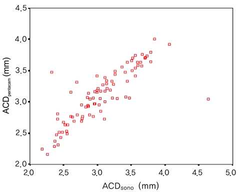
Fig. 2 Correlation of anterior chamber depth (mm) by Pentacam (ACDpentacam) and ultrasound (ACDsono) (r = 0.822, p < 0.05).
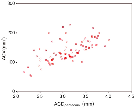
Fig. 3 Correlation between anterior chamber depth (ACD) measured by Pentacam (ACDpentacam) and anterior chamber volume (ACV) (r = 0.650, p = 0.000).
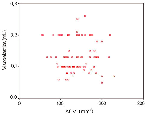
Fig. 4 Correlation between anterior chamber volume (ACV) and injected amount of viscoelastics before capsulorrhexis (r = -0.021, p = 0.850).

Fig. 5 Correlation between anterior chamber depth by Pentacam (ACDpentacam) and injected amount of viscoelastics before capsulorrhexis (r = 0.123, p = 0.253).
Reference
-
1. Ucakhan OO, Gesoglu P, Ozkan M, Kanpolat A. Corneal elevation and thickness in relation to the refractive status measured with the Pentacam Scheimpflug system. J Cataract Refract Surg. 2008. 34:1900–1905.2. Park JY, Kim SY, Jung MS. Comparison of corneal thickness and anterior chamber depth measured with Orbscan, Pentacam, and Ultrasound Pachymetry. J Korean Ophthalmol Soc. 2009. 50:664–669.3. Shin YJ, Kim NH, Kim DH. Comparison of Pentacam with Orbscan. J Korean Ophthalmol Soc. 2007. 48:637–641.4. Kwon SM, Oh HC, Lee DJ, et al. Comparison of anterior segment parameters in angle-closure glaucoma using Scheimpflug camera. J Korean Ophthalmol Soc. 2009. 50:128–134.5. Rabsilber TM, Khoramnia R, Auffarth GU. Anterior chamber measurements using Pentacam rotating Scheimpflug camera. J Cataract Refract Surg. 2006. 32:456–459.6. Nemeth G, Vajas A, Kolozsvari B, et al. Anterior chamber depth measurements in phakic and pseudophakic eyes: Pentacam versus ultrasound device. J Cataract Refract Surg. 2006. 32:1331–1335.7. Pei X, Bao Y, Chen Y, Li X. Correlation of lens density measured using the Pentacam Scheimpflug system with the Lens Opacities Classification System III grading score and visual acuity in age-related nuclear cataract. Br J Ophthalmol. 2008. 92:1471–1475.8. Kubo E, Kumamoto Y, Tsuzuki S, Akagi Y. Axial length, myopia, and the severity of lens opacity at the time of cataract surgery. Arch Ophthalmol. 2006. 124:1586–1590.9. Chylack LT Jr, Wolfe JK, Singer DM, et al. The Lens Opacities Classification System III. The Longitudinal Study of Cataract Study Group. Arch Ophthalmol. 1993. 111:831–836.10. Koranyi G, Lydahl E, Norrby S, Taube M. Anterior chamber depth measurement: a-scan versus optical methods. J Cataract Refract Surg. 2002. 28:243–247.11. Hoffer KJ. Axial dimension of the human cataractous lens. Arch Ophthalmol. 1993. 111:914–918.12. Jivrajka R, Shammas MC, Boenzi T, et al. Variability of axial length, anterior chamber depth, and lens thickness in the cataractous eye. J Cataract Refract Surg. 2008. 34:289–294.13. Walkow T, Anders N, Klebe S. Endothelial cell loss after phacoemulsification: relation to preoperative and intraoperative parameters. J Cataract Refract Surg. 2000. 26:727–732.14. Hayashi K, Hayashi H, Nakao F, Hayashi F. Risk factors for corneal endothelial injury during phacoemulsification. J Cataract Refract Surg. 1996. 22:1079–1084.15. O'Brien PD, Fitzpatrick P, Kilmartin DJ, Beatty S. Risk factors for endothelial cell loss after phacoemulsification surgery by a junior resident. J Cataract Refract Surg. 2004. 30:839–843.16. Cho YK, Kim EC, Kim MS. The surgical results of phacoemulsification cataract surgery according to pupil size. J Korean Ophthalmol Soc. 2007. 48:761–767.17. Gimbel HV. Nucleofractis phacoemulsification through a small pupil. Can J Ophthalmol. 1992. 27:115–119.18. Alio JL, Mulet ME, Shalaby AM, Attia WH. Phacoemulsification in the anterior chamber. J Cataract Refract Surg. 2002. 28:67–75.19. Fine IH, Packer M, Hoffman RS. Steinert RF, editor. Phacoemulsification in the presence of a small pupil. Cataract surgery: technique, complication and management. 2004. 2nd ed. Philadelphia: Saunders;211–222.
- Full Text Links
-
- Actions
-
Cited
- CITED
-
- Close
- Share
-
- Similar articles
-
- Effect of Anterior Chamber Depth on Corneal Endothelial Change After Phacoemulsification
- Endothelial Cell Damage in Microincison Cataract Surgery and Coaxial Phacoemulsification
- Measurement Comparison of Anterior Segment Parameters between AL-Scan(R) and Pentacam(R)
- Influence of Preoperative Corneal Endothelial Status on Postoperative Corneal Endothelium Density after Cataract Surgery
- Comparison of Short-term Corneal Endothelial Changes following Phacoemulsification through a Limbal and Clear Corneal Incision
